
- Health Conditions A-Z
- Health & Wellness
- Nutrition
- Fitness
- Health News
- Ayurveda
- Videos
- Medicine A-Z
- Parenting
Why Do I Feel A Falling Sensation In Sleep?

Have you ever been so tired that the moment you hit the bed, you feel like you are sinking in your mattress? Or did you ever feel your leg twitch in middle of your sleep? Did you feel like you moved your leg to prevent yourself from falling down? If yes, then you are in for a great read!
Why does this happen?
You are not alone, this happens to around 70% of the total population in some way or another. Although these are signs of sleep disorders, it can also happen to people who are otherwise healthy. Sleep disorder specialists believe that there are certain common triggers that leads to such a sensation.
Our brain is exceptional and sometimes when we dream, it makes us believe that we our part of the dream. And if you are falling from somewhere in your dream, chances are, you would feel like you are falling in real life. This experience is known as hypnic jerk, the movement of your muscle that helps you be safe from that fall, or the imaginary one.
What causes it? When we sleep, our bodies become paralyzed, but that does not mean that our muscle control is not totally off. Rather, a slow, dimmer switch. When the dim switch randomly is turned on, we get jerky movements in our sleep.
This involuntary muscle movement is also called myoclonus or hypnic myoclonus, that transitions as you shift from one sleep phase into another.
ALSO SEE: What is sleep paralysis?
What Could Cause It?
There are many theories on what might be causing it. One of them is that when you are at this stage of sleep, you are still not in deep sleep and your brain misinterprets it as wakefulness, however, at the same time your brain does not recognize your muscles to be moving. This is what leads your brain to send a message to your muscles to check-in. It is a way your brain tries to protect you.
Another theory is of course that of stress. When you are stressed, your mind might be racing, while your body is in stationary. This can also affect your sleep cycle and your sleep can be disrupted.
If you are on stimulant drugs or alcohol, it too can prevent you from achieving a good night sleep. If you have consumed too much caffeine then too your muscles can twitch to make you stay awake.
Furthermore, lack of sleep could also be the reason why your mind is used to staying awake even if your body really needs the rest.
Periodic Limb Movement Disorder
While the falling sensation in sleep is quite common, and happens to healthy people, a severe form is the Periodic Limb Movement Disorder (PLMD). PLMD is a repetitive cramping or jerking of legs during sleep. It is a movement disorder that happens only during sleep. It is called "periodic" because the movements are repetitive and rhythmic, which means it occurs in every 20 to 40 seconds. It is also often linked with restless legs syndrome, however, the two are not the same thing.
Restless leg syndrome on the other hand is a condition that involves strange sensations in the legs and arms while awake and an irresistible urge to move the limbs to relive the sensations.
What causes PLMD may be more severe things, including diabetes, iron deficiency, spinal cord tumor, spinal cord injury, sleep apnea, uremia, anemia, narcolepsy, or certain medications.
Can Mental Health Improvement Help Lower High Blood Pressure? Study Points Towards 3 Habits
Credit: AI
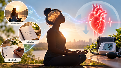
High blood pressure can be managed with medication and dietary changes. But recent research suggests that certain mental health habits can also help fight hypertension.
Mental health practices like mindfulness, optimism and gratitude may also support cardiovascular health, partly by helping people manage stress better and maintain healthier habits.
Hypertension affects nearly half of adults in the United States. While lifestyle changes like reducing salt intake, exercising regularly, maintaining a healthy weight and limiting alcohol remain crucial, the emerging study says that psychological wellbeing can also have a beneficial effect on blood pressure.
“Stress contributes to hypertension,” said Willie E. Lawrence Jr., a cardiologist and spokesperson for the American Heart Association. “It’s hard to get all the stress out of your life, but there are things that you can do to lower blood pressure, like mindfulness.”
According to the researchers, here are three mental health habits that may help:
Mindfulness
Mindfulness refers to paying attention to thoughts, emotions and physical sensations in the present moment without judging them. It may help alleviate stress and improve behaviours that affect blood pressure like dietary choices and physical activity.
Eric Loucks, professor of epidemiology and director of the Mindfulness Center at Brown University, developed a mindfulness-based blood pressure reduction programme that teaches participants all about self-awareness, attention control and emotional regulation. The programme teaches people to apply these skills to everyday decisions like choosing healthier foods.
“There can be energy that comes up to act on those insights,” Loucks said. “So if we decide we want to eat more healthily, can we be there with that energy in the grocery store, selecting the food for the week?”
Optimism
Also read: Eating A Handful Of Nuts Daily May Lower High Blood Pressure Risk, Study Finds
Optimism means expecting positive outcomes while recognising that challenges exist. Research has linked an optimistic outlook with better cardiovascular health, but evidence that it lowers blood pressure in people who already have hypertension is limited.
A study based on more than 100,000 active-duty US Army personnel without hypertension found that the most optimistic participants were less likely to develop high blood pressure during an average follow-up of three and a half years.
Julia Boehm, professor and chair of psychology at Chapman University said that practising optimism does not mean ignoring difficulties. Instead, people can identify realistic, achievable actions that support their health, such as choosing nutritious meals or becoming more physically active.
“This might mean something concrete like, ‘Am I going to choose to eat the salad or the french fries at lunch?’ and thinking optimistically that my choices in any given moment are going to translate into measurable effects in the long run,” Boehm said.
Gratitude
Gratitude involves acknowledging the people, experiences and acts one appreciates. Some research suggests it may affect cardiovascular responses to stress.
One study found that writing a gratitude letter changed how systolic blood pressure responded during a stress test. Systolic pressure, the upper number in a blood pressure reading, measures pressure when the heart contracts.
Mindfulness, optimism and gratitude should complement established hypertension treatment, not replace it. Medication, regular physical activity, a balanced diet and screening remain essential.
As Boehm cautioned, “You can’t ignore the doctor’s advice to either take your medications or engage in physical activity or eat fruits and vegetables. It’s just one piece of a larger strategy to bolster health.”
Long Term Air Pollution Exposure Linked With 14% Higher Dementia Risk: UK Study Based On 488,000 People
Credit: AI
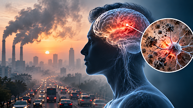
The fine particulate in air pollution could do more damage than brain inflammation. New research indicates long-term exposure to air pollution could lead to changes in the brain structure, rapid biological ageing and a higher risk of dementia.
A study from the UK Biobank, which tracks the health of hundreds of thousands of people in Britain, found that those exposed to the highest levels of fine particulate matter (PM2.5) had a 14% higher risk of developing dementia than those with the lowest exposure.
Researchers examined more than 400,000 participants for nearly 14 years, during which almost 9,000 dementia cases were recorded.
The findings, published in the Journal of Nutrition, Health & Aging in April 2026, also identified that greater pollution exposure could be linked to reduced brain volumes.
Polluted Air Could Affect The Brain
Also read: Heatwaves, Noise & Air Pollution Can Harm Mental Health Too: New Global Commission Warns
PM2.5 consists of particles small enough to penetrate deep into the lungs. Some components can enter the bloodstream, while other particles may reach the brain through ways that are still being investigated.
One possibility is that pollution triggers inflammation throughout the body, which can affect blood vessels and brain tissue. Researchers are also examining whether particles that we inhale can reach the brain directly and influence the proteins, cells and processes that lead to neurodegenerative diseases.
Caleb Finch, professor emeritus at the University of Southern California's Leonard Davis School of Gerontology, said evidence increasingly points towards a relationship between pollution exposure and dementia risk.
“There’s a lot we don’t understand yet, but we do know that the risk of dementia increases in proportion to the amount of air pollution,” he said.
Air Pollution & Dementia
Also read: 1 In 5 US Lung Cancer Patients Never Smoked: Report Highlights Hidden Risk Factors
Earlier research has identified mechanisms that link polluted air with neurological disease.
A study published in Environmental Pollution in May 2026 examined nearly 270,000 people over approximately 14 years. Greater exposure to PM2.5 led to structural changes in the hippocampus, a brain region responsible for learning and memory.
Inflammation and changes in fat metabolism appeared to explain the connection between pollution and dementia.
Another study, published in Science in 2025, examined hospital records from 56.5 million Americans between 2000 and 2014. It found that people living in areas with higher PM2.5 levels were more likely to be hospitalised with dementia with Lewy bodies and Parkinson's disease dementia.
Experiments in mice indicated that pollution exposure triggered abnormal folding and accumulation of alpha-synuclein, a protein that forms the deposits known as Lewy bodies. The animals also developed brain shrinkage, nerve-cell death and cognitive decline.
Xiaobo Mao, a study author and professor of neurology at Johns Hopkins University, said, “This is something we were so surprised to see.”
When researchers exposed mice that were genetically engineered without alpha-synuclein to the same pollution levels, a significant amount of damage was reduced. This suggested that the protein may play a role in the reduction.
Outcomes From Different Airborne Particles
In a separate experiment published in September 2026, researchers exposed mice to different airborne particles like diesel exhaust, dust collected from the World Trade Center site and wood smoke.
Diesel exhaust and World Trade Center dust changed more than 1,200 of the same genes, including genes involved in inflammation and amyloid processing.
Wood smoke did not produce changes related to Alzheimer’s. The researchers observed that diesel exhaust and World Trade Center dust contained more metals, cementing the possibility that certain components of pollution may contribute to neurological harm more than others.
How To Reduce Air Pollution Exposure
Individuals can take a few practical steps to reduce exposure to air pollution. These include improving ventilation when cooking, using appropriate indoor air filtration, replacing heating and ventilation system filters regularly and limiting prolonged exposure to outdoor pollution.
Mao said, “Indoor cooking may sometimes be worse than anything else.” He also emphasised that prevention involves maintaining a balanced diet, exercising and spending time outdoors when air quality is better.
The Altruism Paradox: Helping Others is the Ultimate Act of Self-Care

Credit: AI
An old idea deserves a second look: caring for others may be one of the most powerful ways to care for ourselves.
In traditional Indian homes, we were taught to care for and share with those around us: neighbours, the staff who worked for us, and the wider community we lived in. This was never thought of as charity. It was an everyday part of life, and it carried great value. The community was almost an extension of oneself and one's family.
The idea that the world is one family is not alien to India. The ancient Sanskrit concept of Vasudhaiva Kutumbakam captures it. Over time, as modern life moved faster, this belief slowly faded and self-care took priority. Today, self-care has become almost a global movement, full of advice on exercise, mindfulness, nutrition and stress reduction. Caring and sharing changed form and became "helping others". That was a welcome shift, because it still nudged us beyond our own wellbeing to do good for someone else.
Science Meets Ancient Wisdom
Remarkably, modern science is now bringing us full circle. A growing body of research confirms what philosophers, spiritual traditions and humanitarian organisations have long understood: when we serve others, we nourish ourselves.
The claim that helping others is the ultimate act of self-care may sound paradoxical.
Psychologists call it the "altruistic paradox". Altruism, the selfless concern for the welfare of others, appears at first glance to mean giving away our time, energy and resources. Yet people who volunteer, care, mentor, teach or serve their communities often receive a great deal in return. The rewards are not material. They are better mental, emotional, social and even physical health.
Studies in public health and psychology consistently link volunteering with greater happiness, stronger social connections, a stronger sense of purpose, lower levels of depression and higher life satisfaction. Some longitudinal studies even suggest that regular volunteering is associated with better physical functioning and lower mortality.
Also read: Heatwaves, Noise & Air Pollution Can Harm Mental Health Too: New Global Commission Warns
Why Does It Work?
Part of the answer lies in biology. Acts of kindness stimulate the release of "feel-good" chemicals in the brain, including dopamine, oxytocin and endorphins. This effect is often called the "helper's high".
Helping others also reduces loneliness and builds a sense of belonging, one of our deepest human needs. People who engage in meaningful service tend to report greater optimism, a clearer sense of purpose and more resilience in difficult times.
The benefits are especially significant in later life. Older adults who volunteer regularly report better emotional wellbeing, stronger social ties and less isolation. Retirement can leave a gap in identity and purpose, and service can fill that space with meaning and connection. Altruistic motivation itself seems to amplify these effects.
Also read: 'I Never Recovered From That Crash': US Physicist Recalls Her Years-Long Battle With Long COVID
Service Above Self
Long before science confirmed these findings, humanitarian organisations built their philosophy around this truth. One of the finest examples are volunteer organisations like Rotary International, whose principal motto, "Service Above Self", has inspired generations of volunteers across the world. The motto expresses a simple belief: personal fulfilment and social progress are deeply connected.
Through projects that deliver safe drinking water, promote education, fight disease, build peace, protect the environment and support emotional health, Rotary members enrich societies every day. Behind these efforts are countless hours of selfless work that nurture human bonds, empathy and optimism.
A Remedy for Modern Times
Many of us are struggling with isolation, anxiety and constant tension. Active service is a powerful remedy. Whether we are tutoring a child, comforting an elderly neighbour, supporting someone's mental wellbeing, planting trees or guiding young people, these acts are much more than simple help. They are investments in shared and personal vitality.
Real self-care needs both inward reflection and outward engagement. Lifting those around us also lifts our own spirit. Dedicating ourselves to others gives us meaning, deepens our social ties and anchors us in a larger human family.
Ancient spiritual traditions and contemporary science arrive at the same insight: supporting others does not distract us from our own fulfilment. It is the most reliable foundation for it.
In an age of division, the quiet grace of helping someone else is a profound source of restoration, for our communities and for ourselves.
- Follow Us :


© $2026 Times Horizon Private Limited