
- Health Conditions A-Z
- Health & Wellness
- Nutrition
- Fitness
- Health News
- Ayurveda
- Videos
- Medicine A-Z
- Parenting
Spotting vs Period vs Bleeding: How To Identify
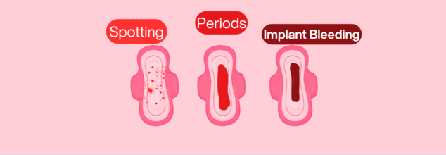
Image Credits: Health and me
A woman's health is intricately linked to her menstrual cycle, which is an important sign of her overall well-being. Throughout puberty and menopause, hormonal changes affect not only fertility but also mood, energy, and long-term health. A normal cycle usually indicates balance, whereas abnormalities may suggest problems such as PCOS, endometriosis, or thyroid disease.
Our bodies do not always work in a perfect clockwork operation and unexpected vaginal bleeding can often confused us. Is it a mere spotting? A normal period? A symptom of something more concerning? Differences between spotting, menstrual bleeding, and intermenstrual bleeding should be understood is crucial for maintaining reproduction health.
Here is a short guide to help you differentiate while you are confused.
Spotting
Spotting is vaginal bleeding that doesn't happen as part of your regular menstrual period. It commonly manifests as fine droplets or smears of blood on clothing or toilet tissue. The intensity of the blood ranges from deep red (recent blood) to pink (having cervical mucus mixed in it) or brown (older, oxidized blood). Spotting is not very much and can't be seen in a way that needs either a tampon or a pad to absorb.
Common Causes of Spotting
Spotting is caused by numerous factors, and in the majority of instances, it is nothing to worry about. Some frequent causes are:
Hormonal Birth Control Transitions: New birth control technique, for instance, birth control pills, IUDs containing hormones, or implants, results in temporary spotting as the body adapts.
Ovulation Bleeding: A few individuals get spotting light around the time of ovulation as a result of hormonal changes. It normally happens in the mid-cycle and could be followed by slight cramping.
Cervical Ectropion: A harmless condition when cells from the inside of the cervical canal migrate to the outer cervix, causing the outer cervix to become more sensitive and prone to faint bleeding on coitus or physical activity.
Early Pregnancy (Implantation Bleeding): 15–25% of pregnant women experience light spotting around 10–14 days post-conception, which is confused with an early period.
When to See a Doctor
Spotting is usually harmless, but it's best to consult a doctor if:
- It continues after a few months of initiating new birth control
- Is accompanied by pelvic pain or abnormal discharge
- Occurs after frequent intercourse
- Occurs during pregnancy and is not verified as implantation bleeding
Menstrual Bleeding
There is a time, also known as a period or menstruation, when the uterine lining sheds due to changing hormone levels. It would last for approximately 2-7 days and is heavier initially. The hue and texture of period blood shift during the menstrual cycle:
Red: New active bleeding at the start of a period
Brown or dark red: Older, slower blood in leaving the uterus
Clots: It's normal to have small clots, but bigger clots may be a sign of heavy menstrual bleeding (HMB)
Why You Have a Period Essentially?
Menstruation is a part of the reproductive cycle, and it happens around every 21–35 days. When there's no pregnancy after ovulation, hormone levels fall, causing the uterine lining to be shed.
Signs Your Period May Be Abnormal
Though periods differ in different people, there are some signs that point towards probable underlying conditions:
- Prolonged bleeding (longer than 7 days)
- Heavy flow necessitating pad/tampon changes every 1–2 hours
- Severe cramps that disrupt daily life
- Irregular periods or absent periods
If you have any of these, conditions such as polycystic ovary syndrome (PCOS), endometriosis, or thyroid disease may be involved, and a medical visit is in order.
Intermenstrual Bleeding
Unlike spotting, intermenstrual bleeding is heavier and unexpected between regular periods. It may be from bright red to dark brown and can contain blood clots.
Possible Causes of Bleeding Between Periods
Sexually Transmitted Infections (STIs): Chlamydia and gonorrhea can lead to inflammation and abnormal bleeding.
Pelvic Inflammatory Disease (PID): A bacterial infection of the reproductive organs and can lead to abnormal bleeding.
Uterine Fibroids or Polyps: Benign growths in the uterus that may cause unexpected bleeding.
Endometrial Hyperplasia or Cancer: In some instances, abnormal bleeding may be a sign of abnormal cell growth in the lining of the uterus.
When to See a Doctor
See a doctor if intermenstrual bleeding is:
- Frequent or persistent
- Associated with pelvic pain or discomfort
- Associated with abnormal discharge, fever or painful sex
Recognizing your body's rhythms can assist you in identifying normal versus abnormal bleeding. Monitoring your menstrual cycle through an app or calendar may flag changes that should be checked with a doctor. If you have any questions regarding abnormal bleeding, visiting your health care provider is the way to go.
Low-Dose Aspirin Shows At Least 70% Lower Dementia Risk In Older Adults But A Serious Complication Still Remains
Credit: iStock
Low-dose aspirin could play a key role in lowering the risk of dementia significantly in some older adults, according to a new genetic analysis. However, the benefit does not come without a complication: major bleeding.
The study, led by researchers at Monash University in Australia, is based on data from more than 13,500 participants in the ASPREE trial, which investigated the effects of low-dose aspirin in older adults. The study was published in Alzheimer’s & Dementia: The Journal of the Alzheimer’s Association.
Researchers found that the benefit was mostly seen among people with a specific genetic trait linked with platelet count.
70% Lower Dementia Risk Was Seen Among Older Adults
Platelets are small blood components that help form clots and stop bleeding. Researchers have previously examined how aggregation of platelets might contribute to dementia, but the role of genetically influenced platelet counts still remains unclear.
The researchers examined genetic scores related to platelet count and compared dementia results among participants who were either assigned aspirin or placebo.
Among people in the highest 20% of platelet count genetic scores, dementia developed in 1% of those taking aspirin, compared with 3.3% of those receiving a placebo. This represented approximately a 70% lower risk.
Lead author Dr Peter Fransquet, a research fellow at Monash University's School of Public Health and Preventive Medicine, said, “Previous trials found aspirin didn't prevent dementia when everyone was considered together.”
“Our findings suggest there may be more to the story,” he said.
Risk Of Bleeding Increased
Also read: Trump Says He ‘Psychologically’ Needs To Pop Aspirin Every Day: But Is It Actually Safe?
The benefit was accompanied by a safety concern. Among participants in the same genetic subgroup, major bleeding occurred in 4.4% of those assigned to aspirin, compared to 2.1% of those given a placebo. That is a difference of 2.3 percentage points in both dementia cases and major bleeding events. In other words, the lower dementia rate cannot be considered in isolation from the increased bleeding risk.
Aspirin reduces the ability of platelets to form clots. This can be useful for certain people who need to take precaution against cardiovascular events, but it can also increase the risk of serious bleeding, including bleeding in the gastrointestinal tract or brain.
The study therefore does not prove that aspirin offers a favourable overall balance of benefits and harms for people with this genetic profile.
Could This Help Personalise Dementia Prevention?
The researchers found that the genetic signal was not linked to established genetic risk factors for Alzheimer's disease and dementia. Instead, it pointed towards platelet factor as a possible direction for further research.
“What's particularly interesting is that the signal wasn't linked to the established genetic risk factors for Alzheimer's disease and dementia,” Fransquet said. “Instead, it's pointing us towards platelet biology and gives us a new avenue to investigate.”
The findings could contribute to more personalised approaches to dementia prevention, in which treatments are tailored to characteristics that influence how individuals respond. But the results need to be replicated in other populations and tested in a clinical trial specifically designed for people with the identified genetic profile.
Fransquet said, “People shouldn’t start taking aspirin to prevent dementia on the basis of this study without consulting with their doctor, particularly given the increased risk of serious bleeding,” he said.
Can Mental Health Improvement Help Lower High Blood Pressure? Study Points Towards 3 Habits
Credit: AI
High blood pressure can be managed with medication and dietary changes. But recent research suggests that certain mental health habits can also help fight hypertension.
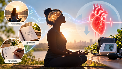
Mental health practices like mindfulness, optimism and gratitude may also support cardiovascular health, partly by helping people manage stress better and maintain healthier habits.
Hypertension affects nearly half of adults in the United States. While lifestyle changes like reducing salt intake, exercising regularly, maintaining a healthy weight and limiting alcohol remain crucial, the emerging study says that psychological wellbeing can also have a beneficial effect on blood pressure.
“Stress contributes to hypertension,” said Willie E. Lawrence Jr., a cardiologist and spokesperson for the American Heart Association. “It’s hard to get all the stress out of your life, but there are things that you can do to lower blood pressure, like mindfulness.”
According to the researchers, here are three mental health habits that may help:
Mindfulness
Mindfulness refers to paying attention to thoughts, emotions and physical sensations in the present moment without judging them. It may help alleviate stress and improve behaviours that affect blood pressure like dietary choices and physical activity.
Eric Loucks, professor of epidemiology and director of the Mindfulness Center at Brown University, developed a mindfulness-based blood pressure reduction programme that teaches participants all about self-awareness, attention control and emotional regulation. The programme teaches people to apply these skills to everyday decisions like choosing healthier foods.
“There can be energy that comes up to act on those insights,” Loucks said. “So if we decide we want to eat more healthily, can we be there with that energy in the grocery store, selecting the food for the week?”
Optimism
Also read: Eating A Handful Of Nuts Daily May Lower High Blood Pressure Risk, Study Finds
Optimism means expecting positive outcomes while recognising that challenges exist. Research has linked an optimistic outlook with better cardiovascular health, but evidence that it lowers blood pressure in people who already have hypertension is limited.
A study based on more than 100,000 active-duty US Army personnel without hypertension found that the most optimistic participants were less likely to develop high blood pressure during an average follow-up of three and a half years.
Julia Boehm, professor and chair of psychology at Chapman University said that practising optimism does not mean ignoring difficulties. Instead, people can identify realistic, achievable actions that support their health, such as choosing nutritious meals or becoming more physically active.
“This might mean something concrete like, ‘Am I going to choose to eat the salad or the french fries at lunch?’ and thinking optimistically that my choices in any given moment are going to translate into measurable effects in the long run,” Boehm said.
Gratitude
Gratitude involves acknowledging the people, experiences and acts one appreciates. Some research suggests it may affect cardiovascular responses to stress.
One study found that writing a gratitude letter changed how systolic blood pressure responded during a stress test. Systolic pressure, the upper number in a blood pressure reading, measures pressure when the heart contracts.
Mindfulness, optimism and gratitude should complement established hypertension treatment, not replace it. Medication, regular physical activity, a balanced diet and screening remain essential.
As Boehm cautioned, “You can’t ignore the doctor’s advice to either take your medications or engage in physical activity or eat fruits and vegetables. It’s just one piece of a larger strategy to bolster health.”
Long Term Air Pollution Exposure Linked With 14% Higher Dementia Risk: UK Study Based On 488,000 People

Credit: AI
The fine particulate in air pollution could do more damage than brain inflammation. New research indicates long-term exposure to air pollution could lead to changes in the brain structure, rapid biological ageing and a higher risk of dementia.
A study from the UK Biobank, which tracks the health of hundreds of thousands of people in Britain, found that those exposed to the highest levels of fine particulate matter (PM2.5) had a 14% higher risk of developing dementia than those with the lowest exposure.
Researchers examined more than 400,000 participants for nearly 14 years, during which almost 9,000 dementia cases were recorded.
The findings, published in the Journal of Nutrition, Health & Aging in April 2026, also identified that greater pollution exposure could be linked to reduced brain volumes.
Polluted Air Could Affect The Brain
Also read: Heatwaves, Noise & Air Pollution Can Harm Mental Health Too: New Global Commission Warns
PM2.5 consists of particles small enough to penetrate deep into the lungs. Some components can enter the bloodstream, while other particles may reach the brain through ways that are still being investigated.
One possibility is that pollution triggers inflammation throughout the body, which can affect blood vessels and brain tissue. Researchers are also examining whether particles that we inhale can reach the brain directly and influence the proteins, cells and processes that lead to neurodegenerative diseases.
Caleb Finch, professor emeritus at the University of Southern California's Leonard Davis School of Gerontology, said evidence increasingly points towards a relationship between pollution exposure and dementia risk.
“There’s a lot we don’t understand yet, but we do know that the risk of dementia increases in proportion to the amount of air pollution,” he said.
Air Pollution & Dementia
Also read: 1 In 5 US Lung Cancer Patients Never Smoked: Report Highlights Hidden Risk Factors
Earlier research has identified mechanisms that link polluted air with neurological disease.
A study published in Environmental Pollution in May 2026 examined nearly 270,000 people over approximately 14 years. Greater exposure to PM2.5 led to structural changes in the hippocampus, a brain region responsible for learning and memory.
Inflammation and changes in fat metabolism appeared to explain the connection between pollution and dementia.
Another study, published in Science in 2025, examined hospital records from 56.5 million Americans between 2000 and 2014. It found that people living in areas with higher PM2.5 levels were more likely to be hospitalised with dementia with Lewy bodies and Parkinson's disease dementia.
Experiments in mice indicated that pollution exposure triggered abnormal folding and accumulation of alpha-synuclein, a protein that forms the deposits known as Lewy bodies. The animals also developed brain shrinkage, nerve-cell death and cognitive decline.
Xiaobo Mao, a study author and professor of neurology at Johns Hopkins University, said, “This is something we were so surprised to see.”
When researchers exposed mice that were genetically engineered without alpha-synuclein to the same pollution levels, a significant amount of damage was reduced. This suggested that the protein may play a role in the reduction.
Outcomes From Different Airborne Particles
In a separate experiment published in September 2026, researchers exposed mice to different airborne particles like diesel exhaust, dust collected from the World Trade Center site and wood smoke.
Diesel exhaust and World Trade Center dust changed more than 1,200 of the same genes, including genes involved in inflammation and amyloid processing.
Wood smoke did not produce changes related to Alzheimer’s. The researchers observed that diesel exhaust and World Trade Center dust contained more metals, cementing the possibility that certain components of pollution may contribute to neurological harm more than others.
How To Reduce Air Pollution Exposure
Individuals can take a few practical steps to reduce exposure to air pollution. These include improving ventilation when cooking, using appropriate indoor air filtration, replacing heating and ventilation system filters regularly and limiting prolonged exposure to outdoor pollution.
Mao said, “Indoor cooking may sometimes be worse than anything else.” He also emphasised that prevention involves maintaining a balanced diet, exercising and spending time outdoors when air quality is better.
- Follow Us :


© $2026 Times Horizon Private Limited