
- Health Conditions A-Z
- Health & Wellness
- Nutrition
- Fitness
- Health News
- Ayurveda
- Videos
- Medicine A-Z
- Parenting
How to Tell If Your Low Blood Pressure Is Dangerously Low? All On Living With Hypotension

How to Tell If Your Low Blood Pressure Is Dangerously Low? All On Living With Hypotension
Low blood pressure, also known as hypotension, is usually considered a sign of good health and low risk for cardiovascular disease. However, there are times when persistently low blood pressure or a sudden drop can be a sign of an underlying health issue that needs medical attention. Knowing when low blood pressure becomes a problem can help ensure timely intervention and proper care.
Blood pressure readings consist of two numbers:
Systolic pressure (the top number) indicates the pressure in your arteries when your heart pumps blood. Diastolic pressure (the bottom number) reflects the pressure in your arteries while your heart is resting between beats.
The normal reading is usually 120/80 mmHg or less. Hypotension is clinically defined as having blood pressure readings less than 90/60 mmHg. In some patients, low blood pressure will have no adverse health consequences and therefore does not need to be treated. In extreme cases, however, it can limit the flow of oxygen and nutrients to vital organs, resulting in potentially life-threatening complications, such as shock.
Causes of Low Blood Pressure
Hypotension can result from many factors. Some of the factors that cause hypotension are as follows:
- Insufficient fluid intake or excessive fluid loss can lead to decreased blood volume and hence hypotension.
- An injury or internal bleeding may result in hypotension. This happens because blood pressure becomes significantly reduced.
- Some hormonal changes along with the increased blood requirement during pregnancy also causes hypotension.
- Issues like heart failure or arrhythmias can impair blood circulation and result in low blood pressure.
- Certain drugs, including diuretics, antidepressants, and blood-pressure-lowering medications, can induce hypotension.
Symptoms of Hypotension
While low blood pressure may not always cause symptoms, it can sometimes be associated with:
- Dizziness or fainting
- Fatigue and weakness
- Blurred vision
- Nausea
- Confusion or difficulty concentrating
- Shallow breathing
- Palpitations
If the person experiences these symptoms, with the recorded blood pressure reading, the medical services have to be consulted.
Types of Low Blood Pressure
1. Orthostatic Hypotension: This is a kind of hypotension that appears as a result of decreased blood pressure when a person is standing up from lying down or sitting position and primarily affects elderly people.
2. Postprandial Hypotension: This is a drop in blood pressure after eating, more common in older people.
3. Neurally Mediated Hypotension: It is triggered by standing for long periods. This type can affect younger people and is associated with miscommunication between the brain and heart.
When Is Low Blood Pressure Dangerous?
There is no such thing as a universal threshold for very low blood pressure, but a blood pressure reading below 90/60 mmHg can be dangerous and require prompt medical assessment if accompanied by symptoms of fainting, confusion, or shortness of breath. Sudden falls in blood pressure may point to potentially serious underlying causes, which can include:
- Internal bleeding
- Sepsis (severe infection)
- Allergic reactions
Management and Treatment
Treatment of hypotension depends on its cause:
- Hydration Drinking more water can help against dehydration-related hypotension.
- Compression Stockings: These can improve circulation and reduce blood pooling in the lower limbs.
- Dietary Changes: Increasing salt intake under supervision can help increase blood pressure
- Medication Changes: The physician may need to reduce dosages of some medications or replace prescriptions.
For more severe cases, physicians might also administer fludrocortisone or midodrine to increase blood pressure. Shock from hypotension should be treated promptly to ensure proper function of organs.
Can Hypotension Cause Fatigue?
Chronic fatigue syndrome has also been associated with low blood pressure. Prolonged tiredness, despite adequate rest, may require reassessment of blood pressure levels. Fatigue secondary to hypotension will impact functionality and thus requires assessment and treatment of the cause.
When to Seek Medical Care
If your blood pressure readings are low consistently and without symptoms, you probably have nothing to worry about. If you do have symptoms like dizziness, fainting, or confusion, you need to go to the doctor. Blood pressure checks are usually conducted regularly. Once you are aware of your own normal baseline, you will know right away when there is something wrong.
Lifestyle Tips That Support Normal Blood Pressure
- Other than directly treating hypotension, a healthy lifestyle overall would promote balanced blood pressure levels.
- Exercise regularly; it promotes cardiovascular health and can help regulate blood pressure.
- Focus on nutrient-dense foods rich in iron, B vitamins, and electrolytes to support circulatory health.
- Excessive alcohol consumption dehydrates the body and lowers blood pressure.
- Practices like yoga and meditation can help maintain a healthy blood pressure range.
Low-Dose Aspirin Shows At Least 70% Lower Dementia Risk In Older Adults But A Serious Complication Still Remains
Credit: iStock
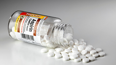
Low-dose aspirin could play a key role in lowering the risk of dementia significantly in some older adults, according to a new genetic analysis. However, the benefit does not come without a complication: major bleeding.
The study, led by researchers at Monash University in Australia, is based on data from more than 13,500 participants in the ASPREE trial, which investigated the effects of low-dose aspirin in older adults. The study was published in Alzheimer’s & Dementia: The Journal of the Alzheimer’s Association.
Researchers found that the benefit was mostly seen among people with a specific genetic trait linked with platelet count.
70% Lower Dementia Risk Was Seen Among Older Adults
Platelets are small blood components that help form clots and stop bleeding. Researchers have previously examined how aggregation of platelets might contribute to dementia, but the role of genetically influenced platelet counts still remains unclear.
The researchers examined genetic scores related to platelet count and compared dementia results among participants who were either assigned aspirin or placebo.
Among people in the highest 20% of platelet count genetic scores, dementia developed in 1% of those taking aspirin, compared with 3.3% of those receiving a placebo. This represented approximately a 70% lower risk.
Lead author Dr Peter Fransquet, a research fellow at Monash University's School of Public Health and Preventive Medicine, said, “Previous trials found aspirin didn't prevent dementia when everyone was considered together.”
“Our findings suggest there may be more to the story,” he said.
Risk Of Bleeding Increased
Also read: Trump Says He ‘Psychologically’ Needs To Pop Aspirin Every Day: But Is It Actually Safe?
The benefit was accompanied by a safety concern. Among participants in the same genetic subgroup, major bleeding occurred in 4.4% of those assigned to aspirin, compared to 2.1% of those given a placebo. That is a difference of 2.3 percentage points in both dementia cases and major bleeding events. In other words, the lower dementia rate cannot be considered in isolation from the increased bleeding risk.
Aspirin reduces the ability of platelets to form clots. This can be useful for certain people who need to take precaution against cardiovascular events, but it can also increase the risk of serious bleeding, including bleeding in the gastrointestinal tract or brain.
The study therefore does not prove that aspirin offers a favourable overall balance of benefits and harms for people with this genetic profile.
Could This Help Personalise Dementia Prevention?
The researchers found that the genetic signal was not linked to established genetic risk factors for Alzheimer's disease and dementia. Instead, it pointed towards platelet factor as a possible direction for further research.
“What's particularly interesting is that the signal wasn't linked to the established genetic risk factors for Alzheimer's disease and dementia,” Fransquet said. “Instead, it's pointing us towards platelet biology and gives us a new avenue to investigate.”
The findings could contribute to more personalised approaches to dementia prevention, in which treatments are tailored to characteristics that influence how individuals respond. But the results need to be replicated in other populations and tested in a clinical trial specifically designed for people with the identified genetic profile.
Fransquet said, “People shouldn’t start taking aspirin to prevent dementia on the basis of this study without consulting with their doctor, particularly given the increased risk of serious bleeding,” he said.
Can Mental Health Improvement Help Lower High Blood Pressure? Study Points Towards 3 Habits
Credit: AI
High blood pressure can be managed with medication and dietary changes. But recent research suggests that certain mental health habits can also help fight hypertension.
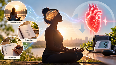
Mental health practices like mindfulness, optimism and gratitude may also support cardiovascular health, partly by helping people manage stress better and maintain healthier habits.
Hypertension affects nearly half of adults in the United States. While lifestyle changes like reducing salt intake, exercising regularly, maintaining a healthy weight and limiting alcohol remain crucial, the emerging study says that psychological wellbeing can also have a beneficial effect on blood pressure.
“Stress contributes to hypertension,” said Willie E. Lawrence Jr., a cardiologist and spokesperson for the American Heart Association. “It’s hard to get all the stress out of your life, but there are things that you can do to lower blood pressure, like mindfulness.”
According to the researchers, here are three mental health habits that may help:
Mindfulness
Mindfulness refers to paying attention to thoughts, emotions and physical sensations in the present moment without judging them. It may help alleviate stress and improve behaviours that affect blood pressure like dietary choices and physical activity.
Eric Loucks, professor of epidemiology and director of the Mindfulness Center at Brown University, developed a mindfulness-based blood pressure reduction programme that teaches participants all about self-awareness, attention control and emotional regulation. The programme teaches people to apply these skills to everyday decisions like choosing healthier foods.
“There can be energy that comes up to act on those insights,” Loucks said. “So if we decide we want to eat more healthily, can we be there with that energy in the grocery store, selecting the food for the week?”
Optimism
Also read: Eating A Handful Of Nuts Daily May Lower High Blood Pressure Risk, Study Finds
Optimism means expecting positive outcomes while recognising that challenges exist. Research has linked an optimistic outlook with better cardiovascular health, but evidence that it lowers blood pressure in people who already have hypertension is limited.
A study based on more than 100,000 active-duty US Army personnel without hypertension found that the most optimistic participants were less likely to develop high blood pressure during an average follow-up of three and a half years.
Julia Boehm, professor and chair of psychology at Chapman University said that practising optimism does not mean ignoring difficulties. Instead, people can identify realistic, achievable actions that support their health, such as choosing nutritious meals or becoming more physically active.
“This might mean something concrete like, ‘Am I going to choose to eat the salad or the french fries at lunch?’ and thinking optimistically that my choices in any given moment are going to translate into measurable effects in the long run,” Boehm said.
Gratitude
Gratitude involves acknowledging the people, experiences and acts one appreciates. Some research suggests it may affect cardiovascular responses to stress.
One study found that writing a gratitude letter changed how systolic blood pressure responded during a stress test. Systolic pressure, the upper number in a blood pressure reading, measures pressure when the heart contracts.
Mindfulness, optimism and gratitude should complement established hypertension treatment, not replace it. Medication, regular physical activity, a balanced diet and screening remain essential.
As Boehm cautioned, “You can’t ignore the doctor’s advice to either take your medications or engage in physical activity or eat fruits and vegetables. It’s just one piece of a larger strategy to bolster health.”
Can Ear Nerve Stimulation Help Lower High Blood Pressure? Scientists Test A New Approach
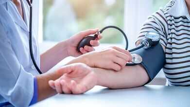
Researchers are testing whether non-invasive nerve stimulation through the ear can help lower blood pressure in people whose hypertension remains uncontrolled despite medication. (Image Credits: iStock)
A small, non-invasive device that stimulates nerves through the ear has shown early promise for people whose blood pressure stays high despite medication, according to a new clinical trial led by researchers at Queen Mary University of London.
What the device does
The device uses a technique called transcutaneous autonomic neuromodulation, or tAN, which aims to influence the autonomic nervous system, the network of nerves that automatically controls functions like heart rate, digestion, and blood pressure, according to Queen Mary University of London. Developed by Afferent Medical Solutions, the handheld device uses two small clips placed on the tragus, the flap of cartilage just in front of the ear canal, to deliver mild electrical pulses. Patients can use it themselves at home without visiting a clinic.
How the trial worked
The trial, called SCRATCH-HTN and funded by the National Institute for Health and Care Research, enrolled 63 people with uncontrolled hypertension, all of whom were already taking at least one blood pressure medication but still had readings above recommended levels, per Queen Mary’s release. Participants were randomly split into two groups: one received active nerve stimulation, the other used a sham device as a comparison.
For the first two weeks, participants used their assigned device for 30 minutes daily. For the following ten weeks, that dropped to once a week.
Also Read: AstraZeneca Cancer Drug Lowers Death Risk By 42% In Advanced Stomach Cancer Trial
What the results showed
The treatment was safe and generally well tolerated, and most participants stuck to the prescribed schedule, suggesting it could work for regular home use, according to the findings published in Hypertension, a journal of the American Heart Association. Across the full study group, however, there was no statistically significant difference in blood pressure between the active stimulation group and the sham group.
There was one notable exception. Among participants who entered the trial with more severe hypertension, those receiving active stimulation showed a greater reduction in blood pressure compared to those on the sham device, per a prespecified subgroup analysis in the study.
What the lead researcher said
“This is an exciting, highly patient-acceptable technology that could ultimately provide a cheap, scalable solution for millions,” said Dr. Ajay Gupta, the trial’s chief investigator and a clinical reader at Queen Mary University of London, in the university’s press release. “Although the trial did not achieve significance across the broad unselected group, those with more severe hypertension exhibited a clear blood pressure reduction in the active group compared to sham.”
He added that the study “points towards important new research directions that could help us understand whether non-invasive neuromodulation can benefit particular groups of patients with uncontrolled hypertension.”
Why this matters
High blood pressure is a major driver of cardiovascular disease, raising the risk of heart attacks and strokes over time. While medication helps many patients, a significant number continue to have blood pressure above target even while on treatment, which is part of why researchers are exploring additional, non-drug approaches that could work alongside existing therapies.
What comes next
The researchers are clear that this is an early finding, not proof of an effective treatment. Because the subgroup result came from a relatively small trial, it will need to be confirmed in larger studies before any conclusions can be drawn about how well the device actually works, and for whom.
- Follow Us :


© $2026 Times Horizon Private Limited