
- Health Conditions A-Z
- Health & Wellness
- Nutrition
- Fitness
- Health News
- Ayurveda
- Videos
- Medicine A-Z
- Parenting
Happy And Safe Diwali: How Smoke From Firecrackers Puts Children's Vision At Risk

Happy And Safe Diwali: How Smoke From Firecrackers Puts Children's Vision At Risk
Diwali is the time of the year when our nation’s children are on holiday and festivities are rampant. Sadly this fun time for children is also when firecrackers and fireworks are available in plenty. The word “firecrackers” evokes quite a bit of emotion and is often associated with great joy but one must realize that the chemical smoke and gases released from firecrackers can be regarded as a serious concern for the capacity of eyes for children.
It is shocking to know that mothers and fathers have been using firecrackers for this long despite it being scientifically proven to be a pollutant and one that can heavily damage children’s eyesight.
But now, as a parent, most people see things differently—firecracker smoke, while dazzling, risks young eyes and health. From choosing eco-friendly lights and indoor activities, ensuring a festive, safe celebration for your children’s well-being is important and necessary. Dr. Neeraj Sanduja, MBBS, MS - Ophthalmology, Ophthalmologist, Eye Surgeon shares all about the toxic effects of smoke from the firecrackers kids so lovingly enjoy bursting and how harmful they can be for their eyes.
Hazardous Effects of Firecracker Smoke
The smoke from firecrackers is a blend of toxic chemicals and fine particles, each carrying potential harm for the delicate tissues of the eyes. Here are some key pollutants present in firecracker smoke that pose specific dangers:
Particulate matter: These are the fumes exhaled from burning that are extremely small but can infiltrate deep into the lungs and eyes creating lesions and scars.
Sulfur dioxide & nitrogen oxides: These gasses emit acid rain, it's easy to comprehend how detrimental it is to the human body.
Heavy metals: If consumed through the skin lead arsenic or any other toxic metals emitted through firecrackers will inflict great damage.
Common Eye Conditions Linked to Firecracker Pollution in Children
Exposure to firecracker smoke can cause a range of immediate symptoms in children, as well as chronic eye problems that may not become apparent until much later. Children are at high risk of developing some symptoms upon exposure to firecracker smoke, and these include:
Red eye syndrome: Clinical eye examinations show that the eyes can be red and watery, as well as itchy due to the smoke's chemicals.
Eye pain: Another common complaint in children is a burning and stinging sensation in the eye areas.
Intolerance to light: The burning of firecrackers leads to the emission of smoke that can irritate the eyes and increase their sensitivity.
Corneal damage: Severe inflammation of the outer layer of the eyeball caused by exposure to smoke may be a more damaging injury caused than previously mentioned damages of the inner layer.
Long-Term Effects of Firecracker Smoke on Eye Health
While some eye issues may appear right after exposure, others may develop subtly and manifest over time, resulting in chronic eye conditions. Dr Neeraj exclaims, "Of great concern is the fact that parents may not notice these initial signs and the signs that are causes are others since these are only some of the diseases that are dangerous but children who continuously come into contact with air polluted by firecrackers may consider the following risks:
Cataracts
What children may notice is a blurriness or cloudiness that gradually spreads but can eventually distort the image fully.Glaucoma
This is a kink in children that often goes unnoticed, where gradual painless deterioration of almost all things alike about vision can be experienced.Dry eye syndrome
However, one chronic problem that can be observed is when there is extreme inflammation in the ocular surface mucus membranes.Tips for Protecting Children’s Eyes During Diwali
Parents can observe the following measures to shield their children's eyes from the harmful effects of firecracker smoke:
Avoid exposure: In the first place, the best way to avoid damaging children’s eyes is to prevent any contact with firecracker smoke. Encourage children to remain in the house or in well-ventilated areas during a firework display.
Wear protective eyewear: In the event that children have to go outside during a firework display, they should wear protective eyewear such as goggles or sunglasses.
Rinse eyes thoroughly: In case of exposure of firework smoke to the child’s eyes, the affected eyes should be washed with plenty of clean water.
Seek medical attention: If a child suffers from intense pain in the eyes, redness in the eyes or any changes in vision after exposure to cracker smoke, he/she should be taken to a medical facility as soon as possible.
Parental Guidance During Diwali
1. Educate Children on Firecracker Safety
2. Monitor Air Quality Index (AQI)
3. Plan Indoor Activities During Peak Pollution Hours
4. Encourage Healthy Eye Habits (Regular Exams, Hygiene)
Eco-Friendly Alternatives to Firecrackers
1. Eco-Friendly Firecrackers
2. Laser Light Shows
3. Community Events (Parades, Festivals)
4. Indoor Games and Activities
5. Environmentally Conscious Celebrations
Firecrackers undoubtedly liven up the festivities; however, the use of firecrackers and their impact on the health of children has caused a lot of concern, especially their vision. It has become crucial to protect young developing eyes from fire cracker pollution which is harmful to them, especially in the long run. In this manner, we are able to assist in the preservation of our children’s eyesight and create safe and happy festive occasions for everyone.
Low-Dose Aspirin Shows At Least 70% Lower Dementia Risk In Older Adults But A Serious Complication Still Remains
Credit: iStock
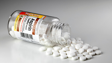
Low-dose aspirin could play a key role in lowering the risk of dementia significantly in some older adults, according to a new genetic analysis. However, the benefit does not come without a complication: major bleeding.
The study, led by researchers at Monash University in Australia, is based on data from more than 13,500 participants in the ASPREE trial, which investigated the effects of low-dose aspirin in older adults. The study was published in Alzheimer’s & Dementia: The Journal of the Alzheimer’s Association.
Researchers found that the benefit was mostly seen among people with a specific genetic trait linked with platelet count.
70% Lower Dementia Risk Was Seen Among Older Adults
Platelets are small blood components that help form clots and stop bleeding. Researchers have previously examined how aggregation of platelets might contribute to dementia, but the role of genetically influenced platelet counts still remains unclear.
The researchers examined genetic scores related to platelet count and compared dementia results among participants who were either assigned aspirin or placebo.
Among people in the highest 20% of platelet count genetic scores, dementia developed in 1% of those taking aspirin, compared with 3.3% of those receiving a placebo. This represented approximately a 70% lower risk.
Lead author Dr Peter Fransquet, a research fellow at Monash University's School of Public Health and Preventive Medicine, said, “Previous trials found aspirin didn't prevent dementia when everyone was considered together.”
“Our findings suggest there may be more to the story,” he said.
Risk Of Bleeding Increased
Also read: Trump Says He ‘Psychologically’ Needs To Pop Aspirin Every Day: But Is It Actually Safe?
The benefit was accompanied by a safety concern. Among participants in the same genetic subgroup, major bleeding occurred in 4.4% of those assigned to aspirin, compared to 2.1% of those given a placebo. That is a difference of 2.3 percentage points in both dementia cases and major bleeding events. In other words, the lower dementia rate cannot be considered in isolation from the increased bleeding risk.
Aspirin reduces the ability of platelets to form clots. This can be useful for certain people who need to take precaution against cardiovascular events, but it can also increase the risk of serious bleeding, including bleeding in the gastrointestinal tract or brain.
The study therefore does not prove that aspirin offers a favourable overall balance of benefits and harms for people with this genetic profile.
Could This Help Personalise Dementia Prevention?
The researchers found that the genetic signal was not linked to established genetic risk factors for Alzheimer's disease and dementia. Instead, it pointed towards platelet factor as a possible direction for further research.
“What's particularly interesting is that the signal wasn't linked to the established genetic risk factors for Alzheimer's disease and dementia,” Fransquet said. “Instead, it's pointing us towards platelet biology and gives us a new avenue to investigate.”
The findings could contribute to more personalised approaches to dementia prevention, in which treatments are tailored to characteristics that influence how individuals respond. But the results need to be replicated in other populations and tested in a clinical trial specifically designed for people with the identified genetic profile.
Fransquet said, “People shouldn’t start taking aspirin to prevent dementia on the basis of this study without consulting with their doctor, particularly given the increased risk of serious bleeding,” he said.
Can Mental Health Improvement Help Lower High Blood Pressure? Study Points Towards 3 Habits
Credit: AI
High blood pressure can be managed with medication and dietary changes. But recent research suggests that certain mental health habits can also help fight hypertension.
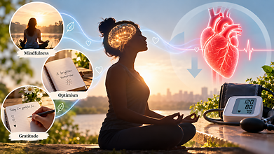
Mental health practices like mindfulness, optimism and gratitude may also support cardiovascular health, partly by helping people manage stress better and maintain healthier habits.
Hypertension affects nearly half of adults in the United States. While lifestyle changes like reducing salt intake, exercising regularly, maintaining a healthy weight and limiting alcohol remain crucial, the emerging study says that psychological wellbeing can also have a beneficial effect on blood pressure.
“Stress contributes to hypertension,” said Willie E. Lawrence Jr., a cardiologist and spokesperson for the American Heart Association. “It’s hard to get all the stress out of your life, but there are things that you can do to lower blood pressure, like mindfulness.”
According to the researchers, here are three mental health habits that may help:
Mindfulness
Mindfulness refers to paying attention to thoughts, emotions and physical sensations in the present moment without judging them. It may help alleviate stress and improve behaviours that affect blood pressure like dietary choices and physical activity.
Eric Loucks, professor of epidemiology and director of the Mindfulness Center at Brown University, developed a mindfulness-based blood pressure reduction programme that teaches participants all about self-awareness, attention control and emotional regulation. The programme teaches people to apply these skills to everyday decisions like choosing healthier foods.
“There can be energy that comes up to act on those insights,” Loucks said. “So if we decide we want to eat more healthily, can we be there with that energy in the grocery store, selecting the food for the week?”
Optimism
Also read: Eating A Handful Of Nuts Daily May Lower High Blood Pressure Risk, Study Finds
Optimism means expecting positive outcomes while recognising that challenges exist. Research has linked an optimistic outlook with better cardiovascular health, but evidence that it lowers blood pressure in people who already have hypertension is limited.
A study based on more than 100,000 active-duty US Army personnel without hypertension found that the most optimistic participants were less likely to develop high blood pressure during an average follow-up of three and a half years.
Julia Boehm, professor and chair of psychology at Chapman University said that practising optimism does not mean ignoring difficulties. Instead, people can identify realistic, achievable actions that support their health, such as choosing nutritious meals or becoming more physically active.
“This might mean something concrete like, ‘Am I going to choose to eat the salad or the french fries at lunch?’ and thinking optimistically that my choices in any given moment are going to translate into measurable effects in the long run,” Boehm said.
Gratitude
Gratitude involves acknowledging the people, experiences and acts one appreciates. Some research suggests it may affect cardiovascular responses to stress.
One study found that writing a gratitude letter changed how systolic blood pressure responded during a stress test. Systolic pressure, the upper number in a blood pressure reading, measures pressure when the heart contracts.
Mindfulness, optimism and gratitude should complement established hypertension treatment, not replace it. Medication, regular physical activity, a balanced diet and screening remain essential.
As Boehm cautioned, “You can’t ignore the doctor’s advice to either take your medications or engage in physical activity or eat fruits and vegetables. It’s just one piece of a larger strategy to bolster health.”
Can Ear Nerve Stimulation Help Lower High Blood Pressure? Scientists Test A New Approach

Researchers are testing whether non-invasive nerve stimulation through the ear can help lower blood pressure in people whose hypertension remains uncontrolled despite medication. (Image Credits: iStock)
A small, non-invasive device that stimulates nerves through the ear has shown early promise for people whose blood pressure stays high despite medication, according to a new clinical trial led by researchers at Queen Mary University of London.
What the device does
The device uses a technique called transcutaneous autonomic neuromodulation, or tAN, which aims to influence the autonomic nervous system, the network of nerves that automatically controls functions like heart rate, digestion, and blood pressure, according to Queen Mary University of London. Developed by Afferent Medical Solutions, the handheld device uses two small clips placed on the tragus, the flap of cartilage just in front of the ear canal, to deliver mild electrical pulses. Patients can use it themselves at home without visiting a clinic.
How the trial worked
The trial, called SCRATCH-HTN and funded by the National Institute for Health and Care Research, enrolled 63 people with uncontrolled hypertension, all of whom were already taking at least one blood pressure medication but still had readings above recommended levels, per Queen Mary’s release. Participants were randomly split into two groups: one received active nerve stimulation, the other used a sham device as a comparison.
For the first two weeks, participants used their assigned device for 30 minutes daily. For the following ten weeks, that dropped to once a week.
Also Read: AstraZeneca Cancer Drug Lowers Death Risk By 42% In Advanced Stomach Cancer Trial
What the results showed
The treatment was safe and generally well tolerated, and most participants stuck to the prescribed schedule, suggesting it could work for regular home use, according to the findings published in Hypertension, a journal of the American Heart Association. Across the full study group, however, there was no statistically significant difference in blood pressure between the active stimulation group and the sham group.
There was one notable exception. Among participants who entered the trial with more severe hypertension, those receiving active stimulation showed a greater reduction in blood pressure compared to those on the sham device, per a prespecified subgroup analysis in the study.
What the lead researcher said
“This is an exciting, highly patient-acceptable technology that could ultimately provide a cheap, scalable solution for millions,” said Dr. Ajay Gupta, the trial’s chief investigator and a clinical reader at Queen Mary University of London, in the university’s press release. “Although the trial did not achieve significance across the broad unselected group, those with more severe hypertension exhibited a clear blood pressure reduction in the active group compared to sham.”
He added that the study “points towards important new research directions that could help us understand whether non-invasive neuromodulation can benefit particular groups of patients with uncontrolled hypertension.”
Why this matters
High blood pressure is a major driver of cardiovascular disease, raising the risk of heart attacks and strokes over time. While medication helps many patients, a significant number continue to have blood pressure above target even while on treatment, which is part of why researchers are exploring additional, non-drug approaches that could work alongside existing therapies.
What comes next
The researchers are clear that this is an early finding, not proof of an effective treatment. Because the subgroup result came from a relatively small trial, it will need to be confirmed in larger studies before any conclusions can be drawn about how well the device actually works, and for whom.
- Follow Us :


© $2026 Times Horizon Private Limited