- Health Conditions A-Z
- Health & Wellness
- Nutrition
- Fitness
- Health News
- Ayurveda
- Videos
- Medicine A-Z
- Parenting
Constant Exposure To Sound May Be Lowering Your Life Expectancy

Credits: Canva
The world that we live in is filled with sounds, some are comforting and some could be jarring too. But what if the constant noise surrounding us is doing us more harm than we realize? There has been immense research that shows that noise is not just a nuisance, but a silent killer and affects our health in ways we do not even know. There are associations of sound causing heart attacks, type 2 diabetes, and dementia.
How Does Your Body Respond To Noise?
Noise is seen as an annoyance, but it effects can go beyond what we imagine. When we hear a sound, it travels through the ear to the brain. This is where it gets processed by the amygdala. It is a region that is responsible for emotional responses. This also triggers a stress response- our heart rate increases, our blood pressure rises, and stress hormones like cortisol flood our system.
This response is also designed to help us react to immediate threats. Especially, if we hear the sound of a predator approaching. However, when we are exposed to constant noise, this response is triggered repeatedly and could compel us to live in a long-term anxious state.
Hidden Health Risks
Many studies including Harvard Health and theAmerican College of Cardiology have found associations of noise with health problems including putting a person at a higher risk of cardiovascular diseases such as heart strokes, attacks, and high blood pressure. The constant activation of the stress response can take a toll on the body, increasing inflammation and making it harder for the heart and circulatory system to function properly. Over time, this can lead to serious health conditions like heart disease and diabetes.
Even more troubling, research suggests that noise pollution may contribute to mental health issues. Studies have found a strong connection between exposure to noise and disturbed sleep, which in turn can cause anxiety, depression, and cognitive decline. The World Health Organization estimates that noise contributes to around 12,000 premature deaths annually across Europe alone. This invisible threat, however, is often overlooked because the effects are gradual and cumulative.
Noise and Sleep: A Silent Disturbance
One of the most insidious aspects of noise pollution is its impact on sleep. Even when we are asleep, our bodies are not fully immune to the effects of sound. Our ears never fully “turn off,” meaning that even faint noises can disrupt our sleep cycle. Research has shown that people who live in noisy environments—whether near busy roads, airports, or urban centers—often experience fragmented sleep, leading to fatigue and a weakened immune system. Over time, this chronic lack of restful sleep can lead to significant health problems, including an increased risk of developing cognitive disorders such as dementia.
The Urbanization Problem
As cities continue to grow, noise pollution is becoming more widespread. Traffic noise, in particular, is one of the most common and harmful sources. The rise of urbanization means more cars, buses, and trains, all of which contribute to an ever-increasing din. This urban soundscape is often relentless, with little respite for those living within it. In densely populated cities, people are exposed to high decibel levels, which can exceed safe thresholds for heart health. In many cases, the sheer volume of sound is not just unpleasant; it’s dangerous.
What Can We Do About It?
The solution is not as simple as reducing noise in our immediate surroundings, though efforts to reduce traffic noise and limit industrial sounds are essential. Some cities have taken steps to create quieter spaces by converting busy roads into pedestrian zones or installing noise barriers. These measures have shown to have a positive impact on public health, with research suggesting that even small reductions in noise can prevent premature deaths and improve overall well-being.
Don't Fear The Biopsy, Fear The Delay

Credit: iStock
For decades, many cancer patients in India have lived with a frightening belief — that a biopsy can make cancer spread. This misconception continues to delay diagnosis, often costing patients the opportunity for timely treatment. This is one of the most common fears I encounter in clinic.
Almost every week, someone tells me, 'Doctor, if you cut it, it will spread.' Families repeat it, neighbors reinforce it, and patients postpone the test. Actually, this concern is linked to a genuine scientific phenomenon known as needle-tract seeding. These phenomena suggest that a few tumor cells may be displaced along the path of the biopsy needle.
However, this event is extremely uncommon and should not be confused with cancer spreading. Seeding is not the same as spreading. A few displaced cells sitting in a needle track are not a metastasis.
Published studies estimate the risk of needle-tract seeding to be extremely low. A 2015 systematic review reported the overall incidence to be below one percent, while more recent reviews, including a 2024 analysis of breast needle biopsy, found no evidence that diagnostic biopsies increase cancer recurrence or reduce long-term survival.
If tumor cells are displaced during a biopsy, they are usually removed when the tumor is surgically excised. In many cases, radiotherapy, systemic treatment and the body's own immune system also eliminate these cells.
In many tier-two and tier-three cities, it takes two to three weeks to reach final report to the treating doctor. During that period, the untreated cancer continues to grow naturally. The patient does not see natural history. He sees cause and effect. The biopsy happened; the lesion grew. The conclusion writes itself — and it is wrong. This delay unintentionally reinforces the misconception that the biopsy triggered the growth, when in reality the cancer was progressing on its own.
At the same time, every tissue injury, including a biopsy, activates the body's wound-healing response, leading to inflammation and new blood vessel formation. Laboratory and animal studies suggest that this temporary inflammatory environment may favor tumor cells. However, these findings have not translated into poorer outcomes for patients. So, the fear is not irrational. It is simply misdirected, and it is mis proportioned.
The diagnosis is not the danger. The delay is. Biopsy helps precision oncology. It helps deciding the treatment approach. Without this we would be treating in the dark.
The consequences of delaying diagnosis are serious. Patients of oral cancer diagnosed while the disease is still localized have a five-year survival rate of nearly 79 per cent. Once the cancer spreads to distant organs, survival drops to around 19 per cent. Unfortunately, nearly two-thirds of oral cancer patients in India are still diagnosed at an advanced stage.
UK Woman Spends £4,000 On Wegovy, Mounjaro, But Loses Less Than 14 Pounds In 15 Months

Credit: iStock/JamPress
A UK woman has claimed that Wegovy and Mounjaro, the blockbuster GLP-1 weight-loss drugs from Novo Nordisk and Eli Lilly, did not help her lose much weight despite more than a year of treatment.
Karen Lay, 56, from Essex, spent £4,000 on the medications, hoping they would help her slim down from 16 stone (224 pounds). However, after 15 months, she lost less than a stone, despite eating very little.
Lay now says the injections were far from a "magic fix" and credits a structured diet for helping her lose two stone, the Daily Mail reported.
Started Wegovy, Mounjaro Out of 'Sheer Desperation'
Lay began taking Wegovy in late 2023 after trying several diets without success. She remained on the medication for nine months, even though she felt something "wasn't right."
“My appetite reduced slightly at first,” Lay, a financial services worker, was quoted as saying. “But not enough to make a meaningful difference, so I increased the dosage each month.”
After seeing limited results with Wegovy, Lay stopped the medication for four weeks before switching to Mounjaro. She described the transition period as experiencing the "worst food noise" and said she gained more than seven pounds while waiting for the first drug to clear from her system.
Lay then spent six months on Mounjaro but said she was "barely eating" and still failed to achieve the weight loss she expected.
“I only lost seven pounds,” she said. “I realised the injections simply weren't effective for me and something had to change.”
Her doctor eventually advised her to stop taking the medication because they were concerned she was not eating enough, the report said.
Lay said the experience was “emotionally exhausting and honestly quite soul-destroying,” and "deeply disheartening.” Watching others succeed on the drugs made it even harder.
Switched to Diet Instead
After discontinuing the injections, Lay adopted a very low-calorie diet with support from expert nutrition advisers who provided personalized guidance.
“Once the medication was fully out of my system, I began to feel genuinely better,” Lay said, adding that her “energy returned, digestion improved, and the constipation disappeared.”
Within a year, she went from a dress size 18 to size 12, the smallest she had been in 17 years, with her weight falling to 13 stone 5 pounds, the report said.
Non-responders To GLP-1 Drugs
Clinical trials have shown that most people taking these medications experience substantial weight loss.
- Wegovy: Average weight loss of about 21% after 72 weeks.
- Mounjaro: Average weight loss of about 22.5% over a similar period.
Around 2.5 million adults in the UK are estimated to be using weight-loss injections, while hundreds of thousands have signed up for the newly approved Wegovy pill, which was rolled out by the NHS this month.
But some people are 'non-responders', meaning they do not lose a meaningful amount of weight despite treatment. Research earlier this year found that around one in 10 people taking GLP-1 medications are considered non-responders.
Why Weight-Loss Drugs May Not Work For All
Although GLP-1 receptor agonists have delivered remarkable results for many people, they do not work the same way for everyone. Obesity is a complex condition influenced by brain signaling, hormones, genetics, and metabolism, meaning treatment responses can vary significantly. Possible reasons include:
- Genetic and hormonal variability
- Underlying medical conditions
- Unrealistic expectations about weight-loss outcomes
- Switching to another GLP-1 medication
- Using older, approved weight-loss medications
- Structured lifestyle changes involving diet and physical activity
- Comprehensive medical management of obesity tailored to the individual.
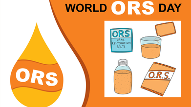
World ORS Day: The Life-Saving Drink Every Home Should Have
Credit: iStock
Oral Rehydration Solution (ORS) is one of the simplest yet most effective life-saving solutions that every household should keep readily available, doctors said on World ORS Day.
Observed every year on July 29, World ORS Day raises awareness about the importance of ORS—a scientifically formulated mix of glucose and electrolytes, including sodium and potassium—that helps prevent and treat dehydration caused by diarrhea, vomiting, fever, heat, and excessive sweating.
First introduced in 1969, ORS has transformed the treatment of dehydration worldwide. Since 2003, the World Health Organization (WHO) and UNICEF have recommended a low-osmolarity ORS formula, which improves fluid absorption and reduces the need for intravenous fluids. Today, ORS remains the gold standard for preventing and treating dehydration, particularly in children and people living in resource-limited settings.
ORS Prevents Dehydration, It Doesn't Treat the Illness
Dr. Ranjana Bhatt, Associate Director - Internal Medicine, Paras Health, Panchkula, told HealthandMe that ORS is not a medicine that cures the underlying illness but is designed to prevent and correct dehydration.
Dehydration occurs when the body loses more water and electrolytes than it takes in, most commonly due to diarrhea or vomiting. It can develop rapidly, especially in young children, older adults, and people with chronic illnesses.
Why ORS Works Better Than Water
ORS works through a well-established biological process in the small intestine. It contains the right balance of glucose and sodium, which are absorbed together through special proteins called sodium-glucose cotransporters (SGLTs). As sodium enters the bloodstream, water follows, allowing the body to rehydrate efficiently.
This mechanism makes ORS more effective than plain water—and often more effective than many commercial sports drinks—for replacing lost fluids and electrolytes quickly and safely.
"Recognizing the early signs of dehydration, such as excessive thirst, dry mouth, dizziness, reduced urination, and unusual fatigue, and starting ORS promptly can prevent serious complications. On ORS Day, we encourage every family to make ORS a household essential and use it appropriately while seeking timely medical care whenever symptoms are severe or persistent," Dr. Bhatt said.
Why Every Household Should Keep ORS
Dr. Amit Prakash Singh, Consultant - Internal Medicine, CK Birla Hospital, Delhi, told HealthandMe that ORS is one of the simplest, most affordable, and most effective ways to prevent dehydration.
"ORS is accessible for almost everyone, and the product is recommended by the World Health Organization to start treating dehydration caused by diarrhea at home. Having ORS packets at home enables treating dehydration without going to the doctor first and potentially avoiding hospitalization," he said.
When Should You Use ORS?
Besides diarrhea, ORS can help replace fluids and electrolytes lost due to:
- Fever-related dehydration
- Vomiting and gastroenteritis
- Heat exhaustion and heatstroke
- Heavy sweating after strenuous exercise
- Hangovers caused by dehydration
- Viral illnesses such as dengue
- Dehydration in older adults and people with chronic illnesses.
"ORS is not just for diarrhea. Whenever there is a lot of fluid loss or electrolyte loss, ORS can be used. ORS can be consumed for vomiting due to gastroenteritis, food poisoning, or viral infection (provided the person can sip small amounts repeatedly)," Dr. Singh said.
ORS Is Not A Cure
Doctors caution that ORS only replaces lost fluids and electrolytes—it does not treat the underlying disease.
It may help prevent dehydration after fever, vomiting, or excessive sweating, but it is not a treatment for abdominal pain, acidity, indigestion, constipation, gastritis, or irritable bowel syndrome (IBS). Persistent or severe symptoms should always be evaluated by a healthcare professional, Dr Singh said.
How To Identify Original ORS
When buying ORS, check the following:
- Look for packaging that states "WHO Recommended Formula." Any other mark or logo may not indicate a WHO-recommended ORS formulation.
- Read the label carefully. Some products may be marketed as "Ready-to-Serve Fruit Beverage" or carry a disclaimer such as: "This is only a trademark and does not represent its true nature. Not a WHO Oral Rehydration Salts." These products are not WHO-recommended ORS.
- Check the ingredients. Genuine ORS should contain glucose, sodium, and potassium in the recommended proportions.
- Follow Us :


© 2024 Bennett, Coleman & Company Limited