- Health Conditions A-Z
- Health & Wellness
- Nutrition
- Fitness
- Health News
- Ayurveda
- Videos
- Medicine A-Z
- Parenting
Can Damaged Sperm Lead To Pregnancy Complications? Study Highlights Uncommon Issues

Can Damaged Sperm Lead To Pregnancy Complications?
Pregnancy is usually a time of happiness and hope, but it also brings in the unexpected. While there is much talk placed on the health of the expectant mother, the quality sperm coming from the father could dramatically change the outcome of pregnancy. The latest study suggests the risks of sperm DNA damage, even increasing the risks of complications preeclampsia and birth prematurity.
In a groundbreaking research study conducted by scientists from Lund University in Sweden, scientists discovered that DNA damage in sperm increases the risk almost up to double that of preeclampsia, this is a dangerous condition that may arise during pregnancies characterized by high blood pressure. In addition, DNA anomalies also increase the risk of premature births, and this further entails increased related adverse health outcomes for infants born through such conditions.
The next step would be to find out which group of men respond best to methods to prevent and treat sperm DNA damage, and to test these methods to prevent pregnancy complications," said Dr. Amelie Stenqvist, a lecturer at Lund University. According to this study, a significant message is put forward that paternal health assumes an important role in a successful pregnancy.
It focused its research on men, specifically whose sperm contained high levels of DNA fragmentation. For instance, some 20% to 30% of babies born via in vitro fertilization have fathers whose sperm contains damaged DNA. The DNA fragmentation index, an indicator to assess the percentage of DNA damage in sperm, indicated that when the percentage of sperm with a DFI above 30% was observed, they had almost no chance of resulting in natural conception. Even a DFI greater than 20% showed that the chances of getting pregnant are highly risky as the risk factor for pregnancy complications like preeclampsia is much high.
Uncommon Complications during Pregnancy
The most alarming complication during pregnancy is preeclampsia. It affects approximately 5% to 8% of pregnancies worldwide, which can cause fatal conditions for both the mother and the baby. The new findings now point out that sperm DNA damage may contribute to this condition, especially if it is due to assisted reproductive techniques such as IVF pregnancies. The research found that a DFI above 20% doubled the risk of preeclampsia from a mere 5% to almost 11% per.
Apart from causing preeclampsia, DNA fragmentation in sperm is also known to increase the risk for prematurity. Most premature babies experience respiratory, neurological, and developmental complications. Therefore, some degree of early intervention might be important for prospective parents.
Some of the rarer, though serious complications include placental abruption, which is the separation of the placenta from the uterine wall and intrauterine growth restriction, a condition by which the baby does not grow normally in the womb. These conditions though rare are potentially catastrophic both to the mother and the child. Results from this study may help in establishing the contribution of the father in such pregnancies.
Further study into sperm DNA damage is of urgent interest with regard to its consequences for pregnancy outcomes. According to Professor Aleksander Giwercman of Lund University in the field of Reproductive Medicine, "the analysis of DFI should be introduced as routine test in all fertility clinics.". "It could give answers to couples who are having difficulties with infertility, but our latest result also shows that DFI analysis can be a method to identify high-risk pregnancies, explained Giwercman.
For many, DNA fragmentation in sperm is often treatable. Common causes are oxidative stress, age, smoking, being obese, and infections. Addressing these elements will likely reduce DNA damage in sperm for men, raising the chances for a healthy pregnancy and baby.
Overall, the study importance should take into consideration paternal as well as maternal health towards reaching for a healthy pregnancy. Though DNA fragmentation in the sperm is supposed to increase the risk factors for complications in pregnancies, the advances into novel treatment approaches and tests are likely to alleviate complications in many families. Thus the findings of this study offer optimism and pave a pathway to more holistic fertility treatments in the future.
How Menopause Changes Heart Health Risks?

Credit: AI
I've spent over three decades treating heart disease, and in that time, I've seen the same misunderstanding come up again and again, across different patients, different generations, different backgrounds. It's the one that delays diagnosis more than any other. Ask most people what menopause changes, and they'll talk about hot flashes, mood swings, or sleep. Ask me, and I'll tell you it changes something just as important: a woman's heart.
I believe menopause should be treated as a cardiovascular milestone, not just a reproductive one. We talk to women a great deal about bone density, hormonal symptoms, and fertility. We talk far less about what's happening quietly inside their arteries during this same period. That gap in the conversation is costing us early diagnoses. In some cases, it's costing lives.
How Estrogen Protects The Heart?
To understand why, it helps to look at what protects a woman's heart in the first place. For most of her reproductive years, estrogen acts almost like a quiet guardian in the background, keeping the endothelium — the inner lining of blood vessels — working well, and supporting the release of nitric oxide, which helps arteries stay relaxed and open. This is a big reason heart attacks are fairly uncommon in women before menopause, compared to men of a similar age.
Menopause takes that buffer away, slowly, not all at once. The shift usually starts during perimenopause, often well before periods actually stop. LDL, the harmful cholesterol, tends to rise; HDL, the protective kind, often falls. Blood pressure that used to be easy to control starts creeping higher. Arteries stiffen, insulin resistance sets in, and fat tends to shift toward the belly, carrying its own inflammatory effect on blood vessels. None of this feels dramatic on any single day. It just builds, quietly, year after year — enough that by ten to fifteen years past menopause, a woman's heart risk has often caught up to, and sometimes overtaken, a man's of the same age.
Also read: Lifestyle Genetics And Hormones: Understanding The Interplay Of Risk Factors For Ovarian Cancer
Ageing & Menopause: A Double Impact
Some argue this is simply age catching up, and that menopause gets blamed unfairly. There's truth in that — age alone stiffens arteries in everyone. But in my experience, losing estrogen compresses those changes into a much shorter stretch of time. It isn't one factor or the other; it's ageing, sped up by a hormonal shift arriving right in the middle of life.
That said, menopause alone doesn't determine a woman's future with heart disease. It sets the stage, but daily habits decide how the story plays out. Smoking, inactivity, weight around the middle, a diet heavy in processed food, unchecked blood pressure and blood sugar, chronic stress, poor sleep, and skipped check-ups all add their share. The reassuring part is that nearly everything on that list can be changed. I've watched patients shift their own trajectory with fairly ordinary steps — walking regularly, eating simply, sleeping better, getting basic tests done once a year.
Also read: Mood Swings, Anxiety & Brain Fog During Menopause? Expert Recommends Looking Beyond Hot Flashes
Heart Symptoms Women Should Never Ignore
What troubles me most in practice is how often real cardiac symptoms get mistaken for "just menopause." Chest pressure rather than sharp pain. New breathlessness during simple activity. Fatigue that doesn't lift with rest. Pain in the jaw, upper back, or between the shoulder blades instead of the classic left-arm pain. Palpitations that don't settle. A few months ago, a woman in her early fifties came to me convinced she had acidity — tightness in the chest, breathlessness on stairs, an occasional fluttering heartbeat. An angiogram showed a significant coronary blockage. "I thought heart problems were a man's disease," she told me. I hear some version of that sentence often, and it worries me every time.
My advice to patients is simple: perimenopause is the right time for an honest conversation with a doctor about the heart, not after something has gone wrong. That means a blood pressure check, a proper lipid profile, blood sugar and HbA1c testing, and a baseline ECG — with an echocardiogram, stress test, or coronary calcium score for those with added risk factors.
Menopause changes a great deal about a woman's body. Her heart is one of those things, whether she's thinking about it or not. Don't wait for a scare to start the conversation. Start it now, while there's still so much you can do.
Don't Fear The Biopsy, Fear The Delay

Credit: iStock
For decades, many cancer patients in India have lived with a frightening belief — that a biopsy can make cancer spread. This misconception continues to delay diagnosis, often costing patients the opportunity for timely treatment. This is one of the most common fears I encounter in clinic.
Almost every week, someone tells me, 'Doctor, if you cut it, it will spread.' Families repeat it, neighbors reinforce it, and patients postpone the test. Actually, this concern is linked to a genuine scientific phenomenon known as needle-tract seeding. These phenomena suggest that a few tumor cells may be displaced along the path of the biopsy needle.
However, this event is extremely uncommon and should not be confused with cancer spreading. Seeding is not the same as spreading. A few displaced cells sitting in a needle track are not a metastasis.
Published studies estimate the risk of needle-tract seeding to be extremely low. A 2015 systematic review reported the overall incidence to be below one percent, while more recent reviews, including a 2024 analysis of breast needle biopsy, found no evidence that diagnostic biopsies increase cancer recurrence or reduce long-term survival.
If tumor cells are displaced during a biopsy, they are usually removed when the tumor is surgically excised. In many cases, radiotherapy, systemic treatment and the body's own immune system also eliminate these cells.
In many tier-two and tier-three cities, it takes two to three weeks to reach final report to the treating doctor. During that period, the untreated cancer continues to grow naturally. The patient does not see natural history. He sees cause and effect. The biopsy happened; the lesion grew. The conclusion writes itself — and it is wrong. This delay unintentionally reinforces the misconception that the biopsy triggered the growth, when in reality the cancer was progressing on its own.
At the same time, every tissue injury, including a biopsy, activates the body's wound-healing response, leading to inflammation and new blood vessel formation. Laboratory and animal studies suggest that this temporary inflammatory environment may favor tumor cells. However, these findings have not translated into poorer outcomes for patients. So, the fear is not irrational. It is simply misdirected, and it is mis proportioned.
The diagnosis is not the danger. The delay is. Biopsy helps precision oncology. It helps deciding the treatment approach. Without this we would be treating in the dark.
The consequences of delaying diagnosis are serious. Patients of oral cancer diagnosed while the disease is still localized have a five-year survival rate of nearly 79 per cent. Once the cancer spreads to distant organs, survival drops to around 19 per cent. Unfortunately, nearly two-thirds of oral cancer patients in India are still diagnosed at an advanced stage.
UK Woman Spends £4,000 On Wegovy, Mounjaro, But Loses Less Than 14 Pounds In 15 Months
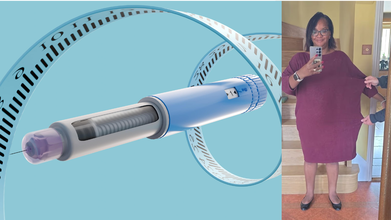
Credit: iStock/JamPress
A UK woman has claimed that Wegovy and Mounjaro, the blockbuster GLP-1 weight-loss drugs from Novo Nordisk and Eli Lilly, did not help her lose much weight despite more than a year of treatment.
Karen Lay, 56, from Essex, spent £4,000 on the medications, hoping they would help her slim down from 16 stone (224 pounds). However, after 15 months, she lost less than a stone, despite eating very little.
Lay now says the injections were far from a "magic fix" and credits a structured diet for helping her lose two stone, the Daily Mail reported.
Started Wegovy, Mounjaro Out of 'Sheer Desperation'
Lay began taking Wegovy in late 2023 after trying several diets without success. She remained on the medication for nine months, even though she felt something "wasn't right."
“My appetite reduced slightly at first,” Lay, a financial services worker, was quoted as saying. “But not enough to make a meaningful difference, so I increased the dosage each month.”
After seeing limited results with Wegovy, Lay stopped the medication for four weeks before switching to Mounjaro. She described the transition period as experiencing the "worst food noise" and said she gained more than seven pounds while waiting for the first drug to clear from her system.
Lay then spent six months on Mounjaro but said she was "barely eating" and still failed to achieve the weight loss she expected.
“I only lost seven pounds,” she said. “I realised the injections simply weren't effective for me and something had to change.”
Her doctor eventually advised her to stop taking the medication because they were concerned she was not eating enough, the report said.
Lay said the experience was “emotionally exhausting and honestly quite soul-destroying,” and "deeply disheartening.” Watching others succeed on the drugs made it even harder.
Switched to Diet Instead
After discontinuing the injections, Lay adopted a very low-calorie diet with support from expert nutrition advisers who provided personalized guidance.
“Once the medication was fully out of my system, I began to feel genuinely better,” Lay said, adding that her “energy returned, digestion improved, and the constipation disappeared.”
Within a year, she went from a dress size 18 to size 12, the smallest she had been in 17 years, with her weight falling to 13 stone 5 pounds, the report said.
Non-responders To GLP-1 Drugs
Clinical trials have shown that most people taking these medications experience substantial weight loss.
- Wegovy: Average weight loss of about 21% after 72 weeks.
- Mounjaro: Average weight loss of about 22.5% over a similar period.
Around 2.5 million adults in the UK are estimated to be using weight-loss injections, while hundreds of thousands have signed up for the newly approved Wegovy pill, which was rolled out by the NHS this month.
But some people are 'non-responders', meaning they do not lose a meaningful amount of weight despite treatment. Research earlier this year found that around one in 10 people taking GLP-1 medications are considered non-responders.
Why Weight-Loss Drugs May Not Work For All
Although GLP-1 receptor agonists have delivered remarkable results for many people, they do not work the same way for everyone. Obesity is a complex condition influenced by brain signaling, hormones, genetics, and metabolism, meaning treatment responses can vary significantly. Possible reasons include:
- Genetic and hormonal variability
- Underlying medical conditions
- Unrealistic expectations about weight-loss outcomes
- Switching to another GLP-1 medication
- Using older, approved weight-loss medications
- Structured lifestyle changes involving diet and physical activity
- Comprehensive medical management of obesity tailored to the individual.
- Follow Us :


© 2024 Bennett, Coleman & Company Limited