- Health Conditions A-Z
- Health & Wellness
- Nutrition
- Fitness
- Health News
- Ayurveda
- Videos
- Medicine A-Z
- Parenting
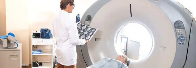
Ultra-Low-Dose CT Scans May Help Early Detection Of Pneumonia
Credit: Canva
Low-dose CT chest scans could help detect pneumonia in at-risk patients while exposing them to only small amounts of radiation, a new study has found. The research, published in Radiology: Cardiothoracic Imaging, shows that ultra-low-dose scans can effectively detect pneumonia in patients with compromised immune systems, enabling doctors to treat the infection before it becomes life-threatening. According to the researchers, these scans expose patients to just 2% of the radiation dose used in a standard CT scan.
"This study paves the way for safer, AI-driven imaging that reduces radiation exposure while preserving diagnostic accuracy,” lead researcher Dr Maximiliano Klug, a radiologist with the Sheba Medical Center in Ramat Gan, Israel, said in a news release. He added that CT scans are the gold standard for detecting pneumonia but there are concerns regarding the risk posed by repeated exposure to radiation. There is a solution- ultra-low-dose CT scan. However, the problem is that these scans can be grainy and hard to read, researchers said.
Study Gives Solution To This
To overcome that, Klug's team developed an AI program that could help "de-noise" low-dose scans, making them sharper and easier to read. Between September 2020 and December 2022, 54 patients with compromised immune systems who had fevers underwent a pair of chest CT scans -- a normal dose scan and an ultra-low-dose scan. The AI program cleaned up the low-dose scan, and then both sets of images were given to a pair of radiologists for assessment. Radiologists had 100% accuracy in detecting pneumonia and other lung problems with the AI-cleaned low-dose scans, but 91% to 98% accuracy in examining the scans that hadn’t been improved through AI, results show.
"This pilot study identified infection with a fraction of the radiation dose," Klug said. "This approach could drive larger studies and ultimately reshape clinical guidelines, making denoised ultra-low dose CT the new standard for young immunocompromised patients.
How Can You Detect Pneumonia?
Pneumonia is a lung infection that causes the air sacs in the lungs to fill with fluid or pus and can be caused by bacteria, viruses, or fungi. The symptoms can range from milk to severe, which includes:
Coughing with or without cough
Fever
Chills
Trouble breathing
Chest pain, especially when breathing deeply or coughing
Sweating or chills
Rapid heart rate
Loss of appetite
Bluish skin, lips, and nails
Confusion.
How to detect Pneumonia in coughing newborns and toddlers?
Pneumonia can severely affect newborns and young children as their lungs are comparatively more sensitive. As per Dr Goyal, young children can cough for various reasons including seasonal infections and tonsillitis, which is very common in this age group. But if they look visibly irritable and have poor sleep patterns, then parents must reach out to an expert. "I am not saying that parents must visit a hospital but any local paediatrician would be able to detect pneumonia in your kid.
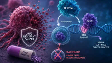
Scientists Find A Previously Unknown Protein That Makes Aggressive Cancers Drug-Resistant
Credit: AI
Scientists have discovered an Achilles' heel that helps some of the most aggressive cancers escape treatment. The discovery that could pave the way for new targeted therapies against specific drug-resistant tumours.
Researchers at Nanyang Technological University (NTU) Singapore, identified a protein called TEX264 that enables cancer cells to escape the therapeutic effects of a widely used class of targeted cancer drugs known as PARP inhibitors. The study is published in Nature Cell Biology.
What Does This Mean For Drug-Resistant Cancers?
The findings could have positive implications for patients with triple-negative breast cancer, as well as ovarian, prostate, and pancreatic cancers that are treated with PARP inhibitors. These cancers eventually become resistant to the drugs.
"Our findings have identified a new biological process involving TEX264 as a key mechanism of drug resistance in an aggressive form of cancer," said Professor Kristijan Ramadan, who led the research. "We coined this new biological mechanism 'autophagy of DNA lesions', or simply 'nucleophagy'. This makes TEX264 a potential target for future cancer therapies."
Also read: Don't Fear The Biopsy, Fear The Delay
How Do Some Cancers Escape Treatment?
PARP inhibitors work by blocking PARP1, a protein that repairs damaged DNA inside cancer cells. Without this repair system, tumour cells accumulate DNA damage and die. However, many cancers eventually become resistant to this treatment.
The researchers discovered that TEX264 helps remove trapped PARP1 proteins from damaged DNA through a newly identified clean-up process called nucleophagy, allowing cancer cells to continue repairing themselves and escape the treatment. This is the cellular recycling system that makes cancer resistant to treatments.
When scientists blocked TEX264 in experiments, PARP1 remained stuck on DNA, DNA damage accumulated, and the cancer cells became far more vulnerable to therapy.
Also read: US FDA Approves New Blood Test To Screen For Colorectal Cancer
What Are PARP Inhibitors?
They are approved for several cancers, including ovarian, breast, prostate, and pancreatic cancer.
While many patients initially respond well, resistance develops in an estimated 40% to 70% of breast and ovarian cancer cases.
Also read: Scientists Find Rare Contagious Skin Cancer In Fish From US, Canada: Should Humans Worry?
Importance Of This Study
Instead of targeting the DNA repair machinery directly, the new study targets an entirely different weak spot, the cellular cycle that cancer cells exploit to discard damaged repair proteins.
Researchers believe drugs that inhibit TEX264 or block nucleophagy could potentially restore the effectiveness of existing PARP inhibitors, offering a new targeted treatment strategy for patients whose cancers no longer respond to therapy.
The researchers caution that the findings are currently based on laboratory studies, and more research, including clinical trials, will be needed before therapies targeting TEX264 can reach patients.
The promising results adds to growing efforts worldwide to overcome one of oncology's biggest challenges: preventing cancers from evolving resistance to life-saving treatments.
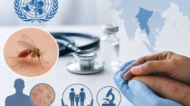
Milestone In Kala-Azar Treatment: WHO Updates Treatment Guidelines, Recommends Shorter & Safer Regimens
Credit: AI
The World Health Organization (WHO) has updated its treatment guidelines for visceral leishmaniasis (VL), commonly known as kala-azar, and post-kala-azar dermal leishmaniasis (PKDL).
Through this update, WHO is recommending shorter, safer and more patient-friendly treatments that could improve outcomes for thousands of patients across eastern Africa and South-East Asia.
The revised guidance marks the first major update in nearly two decades for these forms of leishmaniasis.
It introduces new treatment options, including the use of oral miltefosine in combination therapies, reducing the need for long courses of painful daily injections.
What Is PKDL?
Post-kala-azar dermal leishmaniasis (PKDL) is a skin condition that appears months or years after a patient recovers from visceral leishmaniasis.
Visceral leishmaniasis, also known as kala-azar or black fever, is a life-threatening parasitic disease caused by Leishmania. It is spread by infected female sandflies. It attacks internal organs like the spleen and liver and is almost always fatal if left untreated.
In post-kala-azar, while patients usually feel well, they develop patches, papules or nodules on the skin that harbour the parasite. This allows sandflies to transmit the infection to others.
Early diagnosis and effective treatment of PKDL are therefore considered essential for interrupting transmission and elimination of kala-azar.
Also read: Uganda Declared Ebola-Free As Congo Outbreak Grows To 3,262 Cases, 1,437 Deaths
About The New Treatment Guidelines
WHO now recommends a 14-day regimen combining oral miltefosine with once-daily paromomycin injections for patients with primary visceral leishmaniasis in eastern Africa.
The new approach replaces older sodium stibogluconate-based regimens that required 17 days of treatment, 34 injections, which were often associated with toxicity.
The guidelines also introduce shorter treatment options for PKDL. For patients in eastern Africa and South-East Asia, WHO recommends new combination treatments that substantially shorten duration. It also allows part of the therapy to be completed at home.
Dr Daniel Ngamije Madandi, WHO Director of Malaria and Neglected Tropical Diseases, "For too long, patients suffering from leishmaniasis have endured treatments nearly as punishing as the disease itself. By recommending safer, shorter and more patient-friendly regimens, we are not just improving care; we are accelerating our fight to eliminate this devastating disease and offering renewed hope to communities across Africa and Asia."
Also read: Ebola Scare In The UK After Humanitarian Worker Monitored In London Hospital; Here's what Happened
Milestone In PKDL Treatment
The updated recommendations also revise the safety guidance for miltefosine in line with advice from the WHO Advisory Committee on the Safety of Medicinal Products and introduce allometric dosing to help optimise treatment across different body weights.
Kala-azar is one of the world's deadliest parasitic diseases after malaria. According to WHO, eastern Africa accounted for nearly 79% of global visceral leishmaniasis cases in 2024, with around half of patients being children under the age of 15.
Transmitted through the bite of infected female sandflies, it causes prolonged fever, weight loss, anaemia and enlargement of the spleen and liver. If left untreated, the disease is fatal in up to 95% of cases.
No Junk Food Near Schools! Maharashtra FDA Bans Sale Of Foods & Drinks High In Fat, Salt & Sugar

Credit: AI
Maharashtra Food And Drug Administration (FDA) has taken a significant step towards promoting healthy eating habits in children. On Wednesday, FDA commissioner Tukaram Mundhe announced that foods and drinks high in fat, sugar, and salt should not be sold within 50 metres of school premises.
"Within school premises and up to 50 metres from the school compound, the sale, advertisement and free distribution of high fat, salt and sugar (HFSS) food items is completely banned by the FDA. It will be implemented strictly," Mundhe said.
Why Did Maharashtra FDA Ban The Sale Of HFSS Food Items?
HFSS food items could neither be sold, advertised, nor distributed free of cost within the prescribed limits around schools in Maharashtra anymore.
The recent ban covers popular junk food items like vada pav, samosas, deep-fried snacks, chips, cold drinks, sugary beverages, chocolates and ice cream.
Mundhe said the state government had issued a compliance order on July 27 to enforce food safety and nutritional standards across educational institutions.
He also said that the move would affect nearly two crore students studying in around 1.08 lakh schools in Maharashtra.
The guidelines would apply to all government, government-aided and private schools affiliated to any education board, meaning that no educational institution would be exempt from the ban.
He also said that canteens, midday meal kitchens, food suppliers and hostel kitchens catering to schools must obtain mandatory registration and licenses under food safety regulations.
He added, "No organisation or institution can prepare or supply food to schools, hostels or under the midday meal scheme without registration and a valid license to operate."
He said school principals, headmasters and local authorities would play a key role in implementing the restrictions.
According to the guidelines, the move is applicable for children from 22 months of age up to students studying in Class 12.
Experts React
Also read: Nearly 1.6 Million Dozen Eggs Recalled in US Over Potential Salmonella Contamination: FDA
HealthandMe spoke to Dr Rahul Verma, Director of Paediatrics and Neonatology at Sir H.N. Reliance Foundation Hospital, Mumbai, about the impact this move would have on children's health.
Dr Verma says that rather than viewing the decision only as a ban on junk food, it should be seen as an attempt to reshape children's food environment.
He says, "When healthier choices become the default in schools, children are more likely to develop better eating habits that can continue into adulthood. Reaching over 1.08 lakh schools and nearly 2 crore students from preschool to Class 12 gives this initiative the potential to influence an entire generation at a stage when lifelong dietary preferences are still being formed."
He also said that the policy also goes beyond simply removing HFSS foods. By making FSSAI licensing mandatory for school canteens, introducing regular compliance checks, and encouraging the inclusion of nutritious foods such as fruits, pulses, and whole grains, it creates a structured system that supports healthier eating rather than relying on restrictions alone.
In an X post, Dr. Sivaranjini, a pediatrician, suggested a different approach which seems to be a much simpler way of differentiating between healthy and unhealthy foods.
She wrote, " Front-of-pack label (FOPL) warnings are the simplest and most effective way to help people make better food choices in India. A clear “High in Sugar” or “High in Salt” or "High in Fat" warning takes seconds to understand. No calculations. No nutrition knowledge. Just a simple signal that says, 'Think twice'".
The doctor says that unhealthy, processed foods are often marketed as healthy due to misleading labels and claims on their packaging.
She added, "The problem with systems like Nutri-Score or Health Star Ratings is that they average everything into one score. A product loaded with sugar can still look “healthy” because it has added protein, fiber, or vitamins."
Providing real-life examples, she informed, "A sugary breakfast cereal still gets HIGH IN SUGAR, even if fibre and vitamins are added to it. An instant noodle high in sodium still gets HIGH IN SODIUM even if protein and vitamins are added to it. A packaged fruit juice with excessive free sugars still gets HIGH IN SUGAR, though it's directly from the fruit."
Although the move is a positive step towards protecting children's health, sustained success will depend on whether healthy food remains accessible, affordable, and appealing to children. If children continue to consume highly processed foods outside school or at home, the long-term benefits may be limited.
- Follow Us :


© 2024 Bennett, Coleman & Company Limited