
- Health Conditions A-Z
- Health & Wellness
- Nutrition
- Fitness
- Health News
- Ayurveda
- Videos
- Medicine A-Z
- Parenting
Is 'Sticky Gunk' In Your Brain The Reason Behind Alzheimer's Disease?
Credits: Canva
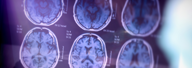
An experimental treatment happens to be the solution to delay Alzheimer's symptoms in some people. These people are the ones who are genetically destined to get the disease in their 40s or 50s. These new findings form ongoing research has now been caught up in Trump administration funding delas. The early results of the study has been published on Wednesday and the participants too are worried that politics could cut their access to a possible lifeline.
One of the participants had said, "It is still a study but it has given me an extension to my life that I never banked on having." The participant is named Jake Henrichs, form New York City, who is 50 years old. He is one of them to be treated in that study for more than a decade now and has remained symptom-free despite inheriting an Alzheimer's-causing gene that had killed his father and brother around the same age.
Slowing Down The Symptoms
Two drugs which can modestly slow down early-stage Alzheimer's are sold in the United States. These drugs clear the brain of one of its hallmarks, a sticky gunk-like part called the amyloid. However, there have not been any hints that removing amyloid far earlier, way many years before the first symptoms appear, may postpone the disease.
How Was The Research Conducted?
The research is led by Washington University in St Louis, which involved families that passed down rare gene mutation as participants. This meant it was almost guaranteed that they will develop symptoms at the same age their affected relatives did.
The new findings is based on a subset of 22 participants who received amyloid-removing drugs the longest, on average eight years. Long-term amyloid removal cut in half their risk of symptom onset. The study is published in the journal Lancet Neurology.
Washington University's Dr Randall Bateman, who directs the Dominantly Inherited Alzheimer's Network of studies involving families with these rare genes says, "What we want to determine over the next five years is how strong is the protection. Will they ever get the symptoms of Alzheimer’s disease if we keep treating them?”
The researchers before though did not know what exactly caused Alzheimer's which affects nearly 7 million Americans, most of them in their later life. However, it is clear that these silent changes occur in the brain at least two decades before the first symptom shows up. The big contributor. At some point amyloid buildup can trigger a protein named tau that then starts to kill neurons, which can lead to cognitive decline.
Researchers are now thus studying the Tau-fighting drugs and are looking into other factors, like inflammation, brain's immune cells and certain virus.
The National Institute of Health (NIH) has expanded its focus as researchers have found more reasons for Alzheimer's. In 2013, the NIH's National Institute on Aging funded 14 trials of possible Alzheimer's drugs over a third targeting amyloid. By last fall, there were 68 drugs and 18% of them target amyloid. However, there are scientists too who think that amyloid is not everything and their is way more in the brain tissue, immune cells, and more which can be studied.
CAR-T Therapy Helps Patients With Rare Autoimmune Disease Walk Again, But Another Trial Reports Death
Credit: AI
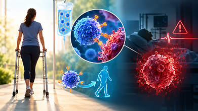
An experimental CAR-T cell treatment has reportedly helped people with stiff person syndrome regain mobility. Some patients even reported not needing walking aids any longer. But new safety concerns have also emerged after Bristol Myers Squibb disclosed a patient's death in a separate early-stage trial of CAR-T therapy for another autoimmune condition, multiple sclerosis.
The developments highlight both the promise and the risks of using these engineered immune cells that are being used to treat autoimmune diseases, rather than cancer.
Science News reported the improvements on October 22, while Reuters reported the death on October 23
CAR-T Therapy Helps Patients With Stiff Person Syndrome Walk Again
Also read: Trump Signs Women’s Health Order: Melania’s 5 Pillars of Innovation Explained
Stiff person syndrome (SPS) is a rare, progressive neurological autoimmune disease that causes severe muscle stiffness, painful spasms and difficulty in walking. Some patients experience sudden spasms that can lead to falls and injuries.
Lynn Montgomery-Haga, who had lived with the condition for about a decade, struggled to walk even short distances and often relied on a walker or wheelchair. After receiving an experimental CAR-T therapy called mivocabtagene autoleucel, or miv-cel, in 2024, her mobility improved significantly.
“I’m just really excited about the future,” Montgomery-Haga told Science News.
Her experience reflects findings from a clinical trial led by neurologist Amanda Piquet at the University of Colorado Anschutz.
One year after treatment, patients continued to show improvements in mobility and disability. Of the 12 participants who needed walking aids during their initial 25-foot walking test, eight completed the follow-up test without one, marking a positive finding.
Piquet told Science News, “You could see that she was walking with a lot more confidence.”
The therapy uses a patient's own T cells, which are modified to recognise and target B cells involved in driving the autoimmune response. The goal is to remove the harmful cells and allow the immune system to rebuild with healthier ones, an approach often described as an “immune reset.”
“It’s revolutionary to think how far things have gone,” said Carl June, a University of Pennsylvania immunologist and oncologist known for his pioneering work on CAR-T cancer treatments.
However, researchers still need longer follow-up and more patient data to understand how reliable the benefits are and what risks may occur.
Bristol Myers Squibb Reports A Patient Death
Also read: 43% Of Women With Autoimmune Diseases Say They Were Told It Was ‘All In Their Head’: Lancet
The safety questions became more pressing after Bristol Myers Squibb disclosed that a patient in its Phase 1 trial of another CAR-T therapy, zolacabtagene autoleucel (zola-cel), died after developing septic pneumonia.
The patient had highly active relapsing multiple sclerosis and developed delayed motor symptoms after receiving the therapy. The symptoms did not respond to treatment. The patient later received high-dose chemotherapy and died from septic pneumonia the following day, 44 days after the CAR-T infusion.
A company spokesperson told Reuters that the patient had “a late onset side effect, a series of other complications and was heavily pretreated prior to CAR T, confounding our ability to determine the root cause of the issue.”
The trial enrolled 66 people with relapsing or progressive forms of multiple sclerosis. Bristol Myers Squibb said most reported side effects were brief and reversible. It also said no additional severe side effects had been reported across its autoimmune CAR-T programmes, which had treated 190 patients in early-stage studies.
The company is working with the US FDA to restart studies, with enrolment targeted to resume in early 2027.
The Findings Need To Be Interpreted Carefully
The two developments involve different CAR-T products and separate clinical trials. The death reported in the zola-cel study does not establish that miv-cel carries the same risk, but it reinforces the need for close safety monitoring.
CAR-T therapy can cause serious complications, including overactive immune system, neurological toxicity and infections. These risks need to be considered when it comes to autoimmune diseases, where patients may otherwise live with their conditions for years and have existing treatment options.
“It’s very important not to oversell it,” said Marc Scherlinger, a rheumatologist at Strasbourg University Hospital, in the Science News report.
AstraZeneca Cancer Drug Lowers Death Risk By 42% In Advanced Stomach Cancer Trial
Credit: AI
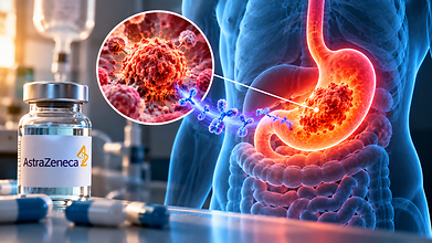
AstraZeneca is developing an experimental cancer drug sonesitatug vedotin that has reported a reduction in the risk of death by 42% in patients with advanced stomach and related cancers. The findings from their Phase 3 clinical trial was presented at the European Society for Medical Oncology (ESMO) Congress in Madrid on October 25.
The global CLARITY-Gastric01 trial compared the drug, also known as sone-ve, to chemotherapy selected by the physician in patients whose cancer had progressed after previous treatment.
The findings showed that patients who received sone-ve lived a median of 10.6 months, compared to 6.4 months among those undergoing chemotherapy.
The trial included 532 patients with advanced gastric cancer, gastroesophageal junction cancer or oesophageal adenocarcinoma whose tumours expressed the protein claudin 18.2 (CLDN18.2). The findings spark hope that the drug could offer another treatment option for patients whose disease has progressed despite early intervention.
Susan Galbraith, AstraZeneca's executive vice-president of oncology haematology research and development, said, “A 5-month improvement in overall survival is very clinically significant in a difficult to treat patient population,” according to Reuters.
How Does Sonesitatug Vedotin Work?
Sonesitatug vedotin belongs to a class of treatments called antibody-drug conjugates (ADCs). These medicines combine an antibody that identifies a target on cancer cells with a drug that kills those cells, becoming a target-focused treatment.
Sone-ve targets CLDN18.2, a protein found on the surface of certain stomach and related cancer cells. The drug is designed to deliver its cancer killing properties to these cells.
The trial enrolled patients whose tumours showed CLDN18.2 expression in at least 25% of tumour cells. This limit could make the treatment relevant to a bigger group of patients than therapies that require higher levels of the protein.
Rui-Hua Xu, professor of medical oncology at Sun Yat-Sen University Cancer Center and the trial's principal investigator, said, “These are remarkable survival results for Sone-Ve in 2nd- and later-line metastatic gastric cancer, a disease where patients face a poor prognosis once initial therapy fails.”
He added that the treatment could be a shift in cancer care for patients whose tumours express CLDN18.2.
Also read: First Human Spinal Cord Transplant Reported In Russia But Can It Reverse Paralysis?
More About The Trial's Findings
The results are promising but the drug did not meet the trial's other primary endpoint, which is survival without disease progression.
Progression-free survival measures how long patients live without their cancer worsening or dying. Median progression-free survival was 4.2 months with sone-ve and 3.8 months with chemotherapy.
Serious side effects related to the treatment occurred in 36.4% of patients receiving sone-ve, compared to 44.8% who did chemotherapy. However, side effects related to stomach were more frequent with sone-ve, affecting 11.1% of patients compared to 6.3% in the control group.
It is important to note that the findings are from a clinical trial and do not mean the drug is approved for routine use. Further regulatory review is needed before it can become a treatment option.
Furthermore, AstraZeneca is also studying sone-ve in early treatment, where it could be used before patients have exhausted multiple therapies.
The company said the findings support investigating the drug in a bigger population of patients with CLDN18.2-positive cancers. The next phase of research will help establish its role in treatment and whether the survival benefit can be maintained when used earlier.
First Human Spinal Cord Transplant Reported In Russia But Can It Reverse Paralysis?
Credit: AI
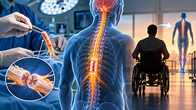
A Chinese-Russian surgical team has reportedly replaced a damaged section of a paralysed patient's spinal cord with tissue from a brain-dead donor, executing the world's first human spinal cord transplant successfully.
However, with no evidence of neurological recovery, scientists have been questioning whether the procedure can restore movement so far. The 12-hour operation was performed at the Sklifosovsky Research Institute for Emergency Medicine in Moscow on October 14–15, according to Chinese surgeon Ren Xiaoping, one of the doctors who participated in the procedure. The patient had sustained a severe spinal cord injury three years ago.
Ren told the South China Morning Post, “It was our Guangxi University of Chinese Medicine Second Affiliated Hospital and the Russian team working together.”
Moscow Mayor Sergei Sobyanin announced the procedure on October 16, describing it as a significant development in spinal surgery. “A patient who was threatened with complete immobility had a chance to recover. Now he feels good,” he wrote.
However, feeling well after surgery does not mean that a patient has regained movement. The surgical team has not publicly released neurological evidence showing whether signals can travel across the transplanted section.
How Was The Spinal Cord Tissue Transplanted?
According to Ren, surgeons removed the damaged section of the patient's spinal cord and replaced it with tissue obtained from a brain-dead donor. They used microsurgery and polyethylene glycol (PEG), a substance that can fuse cell membranes to join the tissues.
This technique addresses one of the central challenges of treating severe spinal cord injuries. The spinal cord carries signals between the brain and the rest of the body. When it is severely damaged, these signals cannot pass normally through the injury, causing paralysis and loss of sensation below the affected area.
But joining two sections of tissue is not enough. Nerve fibres must establish functional connections across the damaged area and the tissue that is transplanted must receive blood supply. The recipient's immune system may also attack the donor tissue.
These challenges become important when the injury is several years old. Repairing a recently damaged spinal cord is different from replacing tissue in a patient with a longstanding injury.
Scientists Have Raised Questions
Also read: Digital Lifestyle and Neck Pain: How to Keep Your Spine Healthy in the Age of Smartphones
Scientists have raised concerns about the evidence about whether it can restore meaningful neurological function in humans.
Melissa Andrews, a neuroscientist at the University of Southampton, questioned the quality of the animal studies supporting the technique and warned about the risks involved, according to New Atlas.
Experiments involving PEG has shown some promise in animals but they do not prove that the technique can reconnect a human spinal cord successfully enough to restore movement.
The lack of publicly available evidence is another limitation. Details about the patient's injury, the precise location of the transplanted segment, neurological examinations after the procedure and changes in movement or sensation have not been disclosed. No peer-reviewed clinical report documenting the operation and its outcomes has been published.
Ren told New Atlas, “We have informed the patient of every potential risk and surgical outcome, and the patient's current condition is good.”
The operation is an experimental treatment, not proof that transplanting spinal cord tissue can reverse paralysis. Even if the patient survives the operation without major complications, recovery would depend on whether functional nerve connections develop and whether the brain can communicate with the body across the damaged region. Rehabilitation and long-term follow-up would be needed to assess any improvement.
- Follow Us :


© $2026 Times Horizon Private Limited