
- Health Conditions A-Z
- Health & Wellness
- Nutrition
- Fitness
- Health News
- Ayurveda
- Videos
- Medicine A-Z
- Parenting
Ebola Outbreak: Uganda Set To Start Vaccine Trials
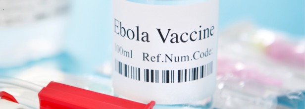
On Thursday, Uganda confirmed an outbreak of the Ebola virus in its capital city Kampala, with the first confirmed patient dying from it a day before. As per the new developments, the officials are now preparing to deploy a trial vaccine to put an end to this outbreak.
Groups of scientists are working on the vaccine and deployment of more than 2,000 doses of a candidate vaccine against the Sudan strain of Ebola has been planned and confirmed by the Uganda Virus Research Institute. As per the World Health Organization (WHO), Uganda has access to 2,169 doses of trial vaccine. For now, however, there are no approved vaccines for the strain and officials are still investigating the source of the outbreak.
The WHO had also allocated $1 million from its contingency fund for emergencies to support quick action and contain the outbreak in the country.
Confirmed Case
On Wednesday, the Sudan strain of Ebola killed a nurse employed at Kampala's main referral hospital. It is after his death that Ebola was declared an outbreak in the country. Post-mortem samples too have confirmed the Sudan Ebola Virus Disease and at least 44 contacts of the deceased man have been listed for tracing. 30 of these are health workers.
Ebola is a highly infectious hemorrhagic fever, which is transmitted through contact with bodily fluids and tissue. Symptoms include headache, vomiting of blood, muscle pains and bleeding.
it was in the late 2022, when Uganda had last suffered an Ebola outbreak. It killed 55 of the 143 people who were infected and was declared over on January 11, 2023.
What Is Ebola Virus Disease?
As per the WHO, Ebola virus disease (EVD) is a rare but severe illness in humans and is often fatal. People can get infected with the virus if they touch an infected animal when preparing food, or touch body fluids of an infected person such as saliva, urine, faeces or semen, or things that have body fluids of an infected person like clothes or sheets.
How Does Transmission Work?
Ebola enters the body through cuts in the skin or when one is touching their eyes, nose or mouth. Early symptoms include fever, fatigue and headache.
It was first discovered in 1976 in two simultaneous outbreak, when in Nzara, South Sudan and other in Yambuku, Democratic Republic of Congo. The latter occurred near a village near the Ebola River, which is where it gets its name from.
It is highly infectious and transmissible disease, in fact, there have been cases of health-care workers who have frequently been infected while treating patients with suspected or confirmed Ebola. This occurs through close contact with patients when infection control precautions are not practiced strictly.
Cases of people conducted burial ceremonies, involving direct contact with the body of the deceased too can lead to the transmission of Ebola. Even after the long suffering and recovery, there is a possibility of sexual transmission. Pregnant women who get acute Ebola and recover may still carry the virus in their breastmilk, or in pregnancy related fluids and tissues.
Symptoms:
- feeling tired
- headache
- muscle and joint pain
- eye pain and vision problems
- weight gain
- belly pain and loss of appetite
- hair loss and skin problems
- trouble sleeping
- memory loss
- hearing loss
- depression and anxiety
AstraZeneca Cancer Drug Lowers Death Risk By 42% In Advanced Stomach Cancer Trial
Credit: AI
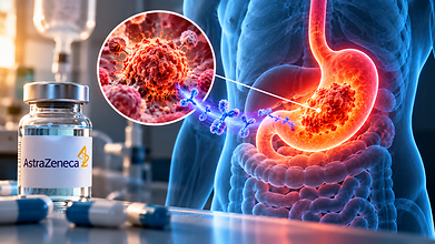
AstraZeneca is developing an experimental cancer drug sonesitatug vedotin that has reported a reduction in the risk of death by 42% in patients with advanced stomach and related cancers. The findings from their Phase 3 clinical trial was presented at the European Society for Medical Oncology (ESMO) Congress in Madrid on October 25.
The global CLARITY-Gastric01 trial compared the drug, also known as sone-ve, to chemotherapy selected by the physician in patients whose cancer had progressed after previous treatment.
The findings showed that patients who received sone-ve lived a median of 10.6 months, compared to 6.4 months among those undergoing chemotherapy.
The trial included 532 patients with advanced gastric cancer, gastroesophageal junction cancer or oesophageal adenocarcinoma whose tumours expressed the protein claudin 18.2 (CLDN18.2). The findings spark hope that the drug could offer another treatment option for patients whose disease has progressed despite early intervention.
Susan Galbraith, AstraZeneca's executive vice-president of oncology haematology research and development, said, “A 5-month improvement in overall survival is very clinically significant in a difficult to treat patient population,” according to Reuters.
How Does Sonesitatug Vedotin Work?
Sonesitatug vedotin belongs to a class of treatments called antibody-drug conjugates (ADCs). These medicines combine an antibody that identifies a target on cancer cells with a drug that kills those cells, becoming a target-focused treatment.
Sone-ve targets CLDN18.2, a protein found on the surface of certain stomach and related cancer cells. The drug is designed to deliver its cancer killing properties to these cells.
The trial enrolled patients whose tumours showed CLDN18.2 expression in at least 25% of tumour cells. This limit could make the treatment relevant to a bigger group of patients than therapies that require higher levels of the protein.
Rui-Hua Xu, professor of medical oncology at Sun Yat-Sen University Cancer Center and the trial's principal investigator, said, “These are remarkable survival results for Sone-Ve in 2nd- and later-line metastatic gastric cancer, a disease where patients face a poor prognosis once initial therapy fails.”
He added that the treatment could be a shift in cancer care for patients whose tumours express CLDN18.2.
Also read: First Human Spinal Cord Transplant Reported In Russia But Can It Reverse Paralysis?
More About The Trial's Findings
The results are promising but the drug did not meet the trial's other primary endpoint, which is survival without disease progression.
Progression-free survival measures how long patients live without their cancer worsening or dying. Median progression-free survival was 4.2 months with sone-ve and 3.8 months with chemotherapy.
Serious side effects related to the treatment occurred in 36.4% of patients receiving sone-ve, compared to 44.8% who did chemotherapy. However, side effects related to stomach were more frequent with sone-ve, affecting 11.1% of patients compared to 6.3% in the control group.
It is important to note that the findings are from a clinical trial and do not mean the drug is approved for routine use. Further regulatory review is needed before it can become a treatment option.
Furthermore, AstraZeneca is also studying sone-ve in early treatment, where it could be used before patients have exhausted multiple therapies.
The company said the findings support investigating the drug in a bigger population of patients with CLDN18.2-positive cancers. The next phase of research will help establish its role in treatment and whether the survival benefit can be maintained when used earlier.
First Human Spinal Cord Transplant Reported In Russia But Can It Reverse Paralysis?
Credit: AI
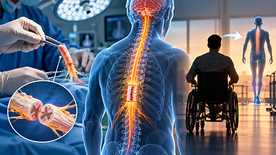
A Chinese-Russian surgical team has reportedly replaced a damaged section of a paralysed patient's spinal cord with tissue from a brain-dead donor, executing the world's first human spinal cord transplant successfully.
However, with no evidence of neurological recovery, scientists have been questioning whether the procedure can restore movement so far. The 12-hour operation was performed at the Sklifosovsky Research Institute for Emergency Medicine in Moscow on October 14–15, according to Chinese surgeon Ren Xiaoping, one of the doctors who participated in the procedure. The patient had sustained a severe spinal cord injury three years ago.
Ren told the South China Morning Post, “It was our Guangxi University of Chinese Medicine Second Affiliated Hospital and the Russian team working together.”
Moscow Mayor Sergei Sobyanin announced the procedure on October 16, describing it as a significant development in spinal surgery. “A patient who was threatened with complete immobility had a chance to recover. Now he feels good,” he wrote.
However, feeling well after surgery does not mean that a patient has regained movement. The surgical team has not publicly released neurological evidence showing whether signals can travel across the transplanted section.
How Was The Spinal Cord Tissue Transplanted?
According to Ren, surgeons removed the damaged section of the patient's spinal cord and replaced it with tissue obtained from a brain-dead donor. They used microsurgery and polyethylene glycol (PEG), a substance that can fuse cell membranes to join the tissues.
This technique addresses one of the central challenges of treating severe spinal cord injuries. The spinal cord carries signals between the brain and the rest of the body. When it is severely damaged, these signals cannot pass normally through the injury, causing paralysis and loss of sensation below the affected area.
But joining two sections of tissue is not enough. Nerve fibres must establish functional connections across the damaged area and the tissue that is transplanted must receive blood supply. The recipient's immune system may also attack the donor tissue.
These challenges become important when the injury is several years old. Repairing a recently damaged spinal cord is different from replacing tissue in a patient with a longstanding injury.
Scientists Have Raised Questions
Also read: Digital Lifestyle and Neck Pain: How to Keep Your Spine Healthy in the Age of Smartphones
Scientists have raised concerns about the evidence about whether it can restore meaningful neurological function in humans.
Melissa Andrews, a neuroscientist at the University of Southampton, questioned the quality of the animal studies supporting the technique and warned about the risks involved, according to New Atlas.
Experiments involving PEG has shown some promise in animals but they do not prove that the technique can reconnect a human spinal cord successfully enough to restore movement.
The lack of publicly available evidence is another limitation. Details about the patient's injury, the precise location of the transplanted segment, neurological examinations after the procedure and changes in movement or sensation have not been disclosed. No peer-reviewed clinical report documenting the operation and its outcomes has been published.
Ren told New Atlas, “We have informed the patient of every potential risk and surgical outcome, and the patient's current condition is good.”
The operation is an experimental treatment, not proof that transplanting spinal cord tissue can reverse paralysis. Even if the patient survives the operation without major complications, recovery would depend on whether functional nerve connections develop and whether the brain can communicate with the body across the damaged region. Rehabilitation and long-term follow-up would be needed to assess any improvement.
From ₹6.2 Lakh To ₹15,900: Kerala Health Minister Defends Generic SMA Drug But Parents Want Safety Assurance
Credit: AI
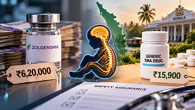
Kerala's decision to switch from an expensive branded medicine for children with spinal muscular atrophy (SMA) with a much cheaper Indian-made alternative has sparked a dispute over the drug’s safety, regulatory procedures and parents' right to know what treatment their children receive.
Health Minister K Muraleedharan has defended the switch to Natco Pharma's generic risdiplam, which is sold as Natsmart. However, some parents and opposition leaders have questioned why bottles supplied to children at the Sree Avittom Thirunal (SAT) Hospital in Thiruvananthapuram were labelled “Physician's Sample” and “For Clinical Trials Only”.
The controversy has since expanded beyond the medicine's price. It now concerns how the drug reached the hospital and what evidence backs its use in children already receiving treatment.
Why Did Kerala Switch To The Generic SMA Medicine?
Also read: 1 In 5 US Lung Cancer Patients Never Smoked: Report Highlights Hidden Risk Factors
SMA is a rare inherited disorder that causes progressive muscle weakness and wasting because of the loss of motor neurons. It can affect movement, swallowing and breathing. Its treatment may need to continue over a long period.
The branded risdiplam, Evrysdi, is manufactured by Swiss pharmaceutical company Roche. Its reported market price is around ₹6.2 lakh per bottle. Natco's generic alternative, Natsmart, is listed at ₹15,900 per bottle. The Kerala government has negotiated even a lower procurement price.
The difference could substantially reduce public expenditure. The state has been providing risdiplam free to eligible children through its KARE programme. Reports indicate that around 109 children are receiving treatment under the scheme.
The generic switch was intended to make treatment more affordable.. Public health expert B Ekbal has backed the decision, arguing that lower-cost medicines can expand access without compromising quality.
The debate is not over whether generic medicines should be used. Opposition leaders, including former chief minister Pinarayi Vijayan, say their objections concern the handling of the medicine that were labelled for clinical trials and the transparency of its distribution.
‘Clinical Trial’ Label Alarmed The Parents
The controversy surfaced after families received bottles bearing the clinical trial and physician sample labels. Parents questioned whether their children had been given medicine intended for testing and whether they had adequately informed before.
Former Kerala health minister and CPI(M) leader K K Shailaja raised concerns about quality and the need for transparency and evidence before changing treatment.
“Just because a new company offers a price reduction doesn’t mean we can purchase everything. Besides price reduction, quality is what matters most. Some companies might lower prices just to push unsold products or for other interests,” she said, according to The South First.
The government says the labels indicate that they belong to sample stock and do not mean that the children were enrolled in a clinical trial.
Dr R Shankar, who heads the technical committee that evaluated the medicine, said, “If the concern is over the label, there was ‘physician’s sample’ written on it. It means it is a tested molecule. The container contains the same medicine.”
Hospital authorities said the disputed bottles were taken back after parents raised concerns. The minister has maintained that this was done to address their apprehensions, not because the government had concluded that the medicine was unsafe.
Muraleedharan has said the switch was based on an expert committee's recommendations and that the drug had received regulatory approval.
On October 24, he said, “People had concerns, but the doctors have addressed them. The treating doctors themselves have cleared the doubts.” He also said he would provide further clarification in the state Assembly on October 26.
Also read: Keralam Senior Citizens Bill: What It Means For India’s Ageing Population
SMA Patient Says The Generic Medicine Worked For Her
Amid the concerns, SMA patient Seba P A has defended Natsmart, saying she has taken Natco's generic risdiplam since April through a patient assistance programme. “Taking Natsmart produces the same effect as when I was taking Evrysdi,” she said. “I have felt no issues due to the change in brand.”
Seba also said bottles supplied to her free of cost carried the same physician sample and clinical trial labels. She argued that the labels were intended to prevent the free medicine from being resold.
Is the Generic SMA Drug Safe?
Natsmart contains risdiplam, the same active pharmaceutical ingredient as Evrysdi. The Central Drugs Standard Control Organisation (CDSCO) has approved Natco's formulation for marketing in India.
They maintain that the generic medicine is suitable for patients and that the labels do not indicate an ongoing clinical trial involving the children.
However, questions have been raised about bioequivalence evidence, quality checks and the process followed before procurement. Bioequivalence studies are used to establish whether a generic formulation delivers an active ingredient into the bloodstream at a rate and to an extent comparable with the reference medicine.
The dispute therefore cannot be resolved by comparing prices or relying solely on individual patient experiences. Families need clear information about the product's approval, the evidence for its use in children, the significance of the labels and the safeguards in place to monitor adverse effects.
- Follow Us :


© $2026 Times Horizon Private Limited